Download
1 / 38
390 likes | 622 Vues
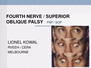
Analysis of Results of Various Surgeries on the Superior Oblique. KOWAL L MAHINDRAKAR A RVEEH, MELBOURNE. Preamble. Superior oblique surgeries are infrequent in Anglo-American strabismus Reputation for being difficult and prone to complications

E N D
Analysis of Results of Various Surgeries on the Superior Oblique KOWAL L MAHINDRAKAR A RVEEH, MELBOURNE
Preamble • Superior oblique surgeries are infrequent in Anglo-American strabismus • Reputation for being difficult and prone to complications • AIM: To examine the results of various superior oblique surgeries (single surgeon)
Methods • Records of 28 patients who had SO surgeries* and who were seen between 2004 -08 were analysed *some had their surgeries outside this period
Diagnostic groups • 1. Superior oblique paresis / palsy SOP • 2. Hypotropia • 3. Brown’s • 4. A pattern
SOP Full tendon width strengthening operation considered if: • Intra- op FDT shows significant unequivocal floppiness [‘..just keeps going’] • Usually: Coronal scan shows atrophy Usual operation: advancement / plication @ insertion till mild Brown’s created [can elevate 6 o’clock limbus ~3mm above the intercanthal horizon]
Results – 4 pts with SO plication • 2 unilateral cong. SOP • 1 bilateral cong. SOP • 1 bilateral acquired SOP due to post. fossa lesion
Unilateral Cong. SOP n=2 • Both had SO plication and Parks’ IO recess • One required re-plication • Both have improved AHP 3 yrs later • One has good ROSV with 100” stereo
Bilateral cong SOP n=1 • Bilateral SO plication • Has large ROSV at 1 year • Torsion corrected from 10° excyclo to < 5° incyclo • Minimal residual hypertropia in R gaze (pre-op 20∆); none in L gaze (pre-op 12∆)
Bilateral SOP 2ary to post. fossa lesion n=1 • Unilateral plication + bilateral IO recession + vertical R-R OU [!] • 13 years follow-up: good ROSV • SO plication corrected 10° of excyclotorsion (8° residual)
Plication for SOP • In selected cases, a reliable and safe operation
Diagnostic groups • 1. Superior oblique paresis / palsy SOP • 2. Hypotropia • 3. Brown’s • 4. A pattern
Hypotropian=4 1-2y follow-up n = 2 Tenotomy • corrected hypo (7∆ and 15∆) • improved A pattern from 25 to 12 ∆ in one pt.
Hypotropian=4 1-2y follow-up n =2: Recession – [posterior] transposition to 13mm from limbus & 2mm nasal to SR (Souza Dias) • corrected hypo (15 and 14∆) • improved A pattern from 25 ∆ to < 5∆
Diagnostic groups • 1. Superior oblique paresis / palsy SOP • 2. Hypotropia • 3. Brown’s • 4. A pattern
Results - Brown’s • 6 patients • Spacer, Tenotomy, Recession • 1-2 years follow-up
Results - Brown’s • Spacer →no improvement (1 pt.) FDT was negative • SO tenotomy + IO recession →no improvement (1 pt.) • SO tenotomy + IO Rc + LR Rc→ Good range of movement (1pt.)
Results - Brown’s • SO tenotomy + IO rec. + other SR faden → improved appearance (1 pt.) • SO spacer (later removed) + IO rec. + other SR faden → No AHP; large ROSV (1 pt.) • SO recession ~12mm → corrected AHP (1 pt.)
Brown’s • These results are not as good as published series • Pathology may vary • 2ary effects [eg tight verticals] may come to dominate the clinical picture and obscure the original pathology
Diagnostic groups • 1. Superior oblique paresis / palsy SOP • 2. Hypotropia • 3. Brown’s • 4. A pattern
A pattern n=14 • 10 : Partial tendon weakening operation : bilateral SO posterior tenectomy [SOPT] • 4 : full tendon width weakening operation 1 full tendon width tenotomy 1 spacer 2 recession – transposition 1-3 y follow-up
Posterior tenectomy NOTE: WIDE SCATTER OF RESULTS From Souza Dias & Praeto Diaz
Posterior tenectomy NOTE: WIDE SCATTER OF RESULTS From Souza Dias & Praeto Diaz
SO Post. Tenectomy n=10 • 1-4 years follow-up • 2/10 patients improved completely • Max. correction 20 ∆
SO Post. tenectomy • 3/10 patients had partial correction • Improvement varied from 10 – 12 ∆ 5/10 had partial or complete correction
SO Post. tenectomy • 3 patients had no improvement • 2 patients : A pattern worse
‘A’ pattern – complete tenotomy n=1 • 2 years follow-up • ‘A’ pattern improved from 25 ∆ to 12 ∆ • Also corrected hypotropia of 7 ∆
‘A’ Pattern – SO spacer n=1 • 1 year follow-up • AHP resolved
‘A’ Pattern Recession – Transposition n=2 • One had correction of 70 ∆ !! • One had improvement in ‘A’ from 25 ∆ to < 5 ∆ + correction of 14 ∆ hypo
Recession - transposition FAIRLY TIGHT RESULTS
SO surgeries • One abandoned as no tendon was found
Complications • One spacer: chronic inflammation and recurrence of Brown’s after temporary improvement • No lid problems
Conclusions • Palsy → plication reliable [if well selected] • Hypotropia → good results from tenotomy, recess / transpose • ‘A’ pattern → Full tendon width procedure better than PTSO • PTSO → not reliable for ‘A’ pattern [but will not ‘bite’ you] • Brown’s → mixed results