Download
1 / 21
260 likes | 1.46k Vues
Globular proteins Myoglobin and hemoglobin. Lecture 5 Dr. Mamoun Ahram Summer, 2014. Functions of myoglobin and hemoglobin. Myoglobin is storage of O 2 in muscles. During periods of oxygen deprivation, oxymyoglobin releases its bound oxygen. Hemoglobin: transport of O 2 and CO 2

E N D
Globular proteinsMyoglobin and hemoglobin Lecture 5 Dr. Mamoun Ahram Summer, 2014
Functions of myoglobin and hemoglobin • Myoglobin is storage of O2 in muscles. During periods of oxygen deprivation, oxymyoglobin releases its bound oxygen. • Hemoglobin: • transport of O2 and CO2 • blood buffering
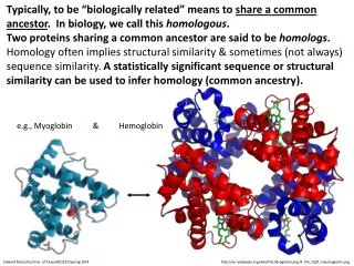
Structure of myoglobin • Myoglobin is a monomeric protein that is mainly found in in muscle tissue. • It includes a prosthetic group, the heme group • It can be present in two forms: • oxymyoglobin (oxygen-bound) • deoxymyoglobin (oxygen-free) • The tertiary structure of myoglobin 8 α-helices, designated A through H, that are connected by short non-helical regions.
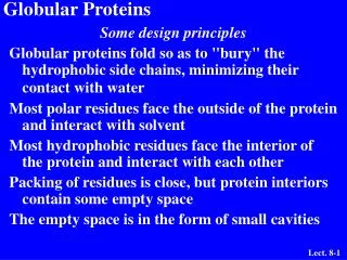
Arrangement of amino acids • Like other globular protein, amino acid R-groups exposed on the surface of the molecule are generally hydrophilic, while those in the interior are predominantly hydrophobic. • Except for two histidine residues in helices E and F (known as E7 and F8) • F8 His is designated as proximal His, whereas E7 His is known as distal His
Heme group • Both proteins contain a heme group. • It is a prosthetic group (a non-protein group covalently attached to a protein). • Heme is a flat molecule that has four cyclic groups known as pyrrole rings.
Iron • Iron can bind in the center of the four rings. • Fe is in the ferrous state (Fe2+) can form 6 bonds: • 4 with the nitrogen of the rings, • One (known as the fifth coordinate) with the nitrogen of a histidine imidazole (known as proximal His). • One with O2 (the sixth coordinate) • Oxidation of iron to the Fe3+, ferric, state makes the molecule incapable of normal O2 binding • Upon absorption of light, heme gives a deep red color.
Structure-function relationship • The planar heme group fits into a hydrophobic pocket of the protein and the myoglobin-heme interaction is stabilized by hydrophobic attractions. • The heme group stabilizes the tertiary structure of myoglobin. • The distal histidine acts as a gate that opens and closes as O2 enters the hydrophobic pocket to bind to the heme. • The hydrophobic interior of myoglobin (or hemoglobin) prevents the oxidation of iron, and so when O2 is released, the iron remains in the Fe(II) state and can bind another O2.
Oxygen binding to myoglobin • Myoglobin binds O2 with high affinity. • The P50 (oxygen partial pressure required for 50% of all myoglobin molecules) for myoglobin ~2.8 torrs or mm Hg • Given that O2 pressure in tissues is normally 20-30 mm Hg, it is almost fully saturated with oxygen at normal conditions. The binding of O2 to myoglobin follows a hyperbolic saturation curve.
Hemoglobinstructure • Hemoglobin is tetrameric hemeprotein (fur protein chains known as globins with each bound to heme. • In adults, the four globin proteins are of two different types known as α and β, so a hemoglobin protein is an α2β2 globin protein. • The α and β chains contain multiple α-helices where α contains 7 α-helices and β contains 8 α-helices (similar to myoglobin).
Chain interaction • The chains interact with each other via hydrophobic interactions • Therefore, hydrophobic amino acids are not only present in the interior of the protein chains, but also on the surface. • Electrostatic interactions (salt bridges) and hydrogen bonds also exist between the two different chains.
Oxygen binding to hemoglobin • Hemoglobin must bind oxygen efficiently and become saturated at the high oxygen pressure found in lungs (approximately 100 mm Hg) • Then, it releases oxygen and become unsaturated in tissues where the oxygen pressure is low (about 30 mm Hg)
The saturation curve • The saturation curve of hemoglobin binding to O2 has a sigmoidal shape • At 100 mm Hg, hemoglobin is 95-98% saturated (oxyhemoglobin) • As the oxygen pressure falls, oxygen is released to the cells • In contrast to a low p50 for myoglobin, the p50 of hemoglobin is approximately 26 mm.
The two structures of hemoglobin High affinity state Transition state Peripheral tissues Lungs Low affinity state
Hemoglobin is allosteric • Hemoglobin is an allosteric protein (from Greek "allos" = "other", and "stereos" = "shape"). • An allosteric protein: a protein where binding of a molecule (ligand) to one part of the protein affects binding of a similar or a different ligand to another part of the protein. • Hemoglobin exists in two forms, T-state and R-state • The T-state is also known as the "taut" or "tense" state and it has a low-binding affinity to oxygen. • The R-state is known as the "relaxed" state and it has 500 times higher affinity to oxygen than as the T conformation . • Binding of O2 causes conformational changes in hemoglobin, converting it from the low affinity T-state to the high affinity R-state .
How does the structure change? (1) • When heme is free of oxygen, it has a domed structure and iron is outside the plane of the heme group. • When oxygen binds to an iron atom, heme adopts a planar structure and the iron moves into the plane of the heme pulling proximal histidine (F8) along with it.
How does the structure change? (2) • This movement triggers • changes in tertiary structure of individual hemoglobin subunits and • breakage of the electrostatic bonds at the other oxygen-free hemoglobin chains.
Not in myoglobin • In myoglobin, movement of the helix does not affect the function of the protein.
The saturation curve is sigmoidal because… • Conformational changes lead to cooperativity among binding sites. • Binding of the first O2 breaks some salt bridges with the other chains increasing the affinity of the binding of a second molecule. • Binding of the second O2 molecule breaks more salt bridges increasing the affinity towards binding of a third O2 even more, and so on.