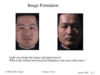
Understanding Visual Image Formation in the Human Eye
This schematic illustrates how visual images are formed in the human eye. Light rays from an object are focused by the cornea and lens to create an inverted image on the retina, located at the back of the eye. The retina contains the fovea, the central spot where photosensitive cells are densely packed, crucial for sharp vision. The optic nerve, connecting to the bright yellow area of the retina, carries visual information to the brain, where it is processed. This image is contributed by Alexander Churkin and is licensed under the GNU Free Documentation License.

Understanding Visual Image Formation in the Human Eye
E N D
Presentation Transcript
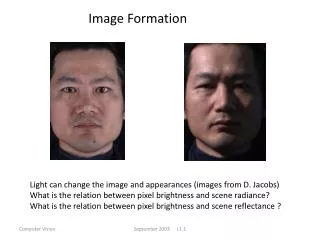
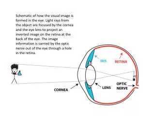
Image formation Schematic of how the visual image is formed in the eye. Light rays from the object are focused by the cornea and the eye lens to project an inverted image on the retina at the back of the eye. The image information is carried by the optic nerve out of the eye through a hole in the retina.
retina Photograph of a human retina. The dark spot in the center of the image is the fovea, where the photosensitive cells are most dense. The optic nerve connects at the bright yellow area on the right, where the blood vessels converge, and where there are no photosensitive cells. Image made available by Alexander Churkin via Wikimedia Commons under the GNU Free Documentation License.