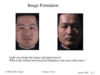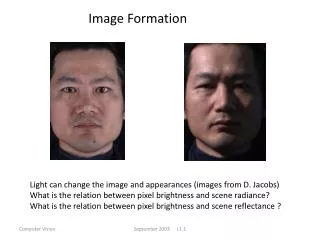
Image Formation
Image Formation. 1-Placing the patient inside the magnet leads to magnetize the tissues protons where they aligned parallel or anti-parallel to the magnetic fieldB0. At this stage the proton precess randomly or out of phase *.


Image Formation
E N D
Presentation Transcript
1-Placing the patient inside the magnet leads to magnetize the tissues protons where they aligned parallel or anti-parallel to the magnetic fieldB0. At this stage the proton precess randomly or out of phase * • *At this stage the proton precess randomly or out of phase for various reasons mainly a- external reasons; the effects of the static strong magnet. b-internal reasons; the effects of the inter proton magnetic interaction. Some facts about the magnetizing the protons: 1-The number of parallel aligned protons increases as the magnet strength increases. 2- As the number of aligned protons increases the MRI signal will be stronger and easily detectable. 3- As the magnet strength increase the resonance frequency(Larmor frequency) increases as well
Steps of imaging • 2- Flip the protons magnetic field from the Bo direction(*) to the XY direction. We do this by sending radiofrequency energy with specific frequency to supply the protons with enough energy to be excited and flip to the transverse (XY) direction.
Steps of imaging • 3- put the protons to precess in phase to resonate and release the absorbed energy as MRI signal. We do this by using step 2; sending the RF will flip the protons and get them in phase as well ( although they stay in phase for very short time)
FID decays exponentially • exponentially means the rate of change either increase or decrease must be expressed using exponents. A graph of such a rate would appear as a curve that continually becomes steeper or shallower, not as a straight line( linear)
The current situation • The protons are out of phase!!!!! • The FID signal was unsuitable to produce an image • We need the protons to be in phase so they resonate and give signal
Tau is the time from the 90° pulse to the rephasing 180° RF pulse • TE =2xTau is the time from the 90° pulse to the Echo
TR is an important factor of imaging, so we need to remember TR
4- Receive the MRI signal. We do this by placing a receiver coil (antenna) in the path of the changing magnetic field
PROTONS MAGNETS • This is faraday’s law of induction: Protons( tiny magnets) flip directions in side the coil induce current or MR signal