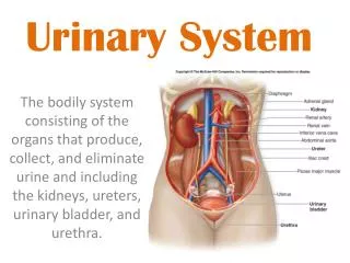
URINARY SYSTEM: II
URINARY SYSTEM: II. (cont.). URINARY SYSTEM: II. TO IDENTIFY THE COMPONENTS OF THE URINARY SYSTEM. TO CHARACTERIZE THE GENERAL ORGANIZATION OF THE KIDNEY. TO EXAMINE THE HISTOLOGICAL STRUCTURE OF THE NEPHRON AND THE COLLECTING DUCTS.

URINARY SYSTEM: II
E N D
Presentation Transcript
URINARY SYSTEM: II (cont.)
URINARY SYSTEM: II TO IDENTIFY THE COMPONENTS OF THE URINARY SYSTEM TO CHARACTERIZE THE GENERAL ORGANIZATION OF THE KIDNEY TO EXAMINE THE HISTOLOGICAL STRUCTURE OF THE NEPHRON AND THE COLLECTING DUCTS TO CORRELATE STRUCTURE OF VARIOUS COMPONENTS WITH FUNCTION
P P P P P RC P P URINARY SYSTEM KIDNEY (ORGANIZATION) CORTEX - region immediately beneath renal capsule - composed of two distinct regions: (1) CORTICAL LABYRINTH (2) MEDULLARY RAY MEDULLA - located immediately beneath renal cortex - consists of triangular blocks of tissue called the PYRAMIDS - RENAL COLUMNS are strands of cortical tissue that extend down between adjacent pyramids
Cortical Labyrinth with interdigitating Medullary Rays RENAL LOBE - a single pyramid with its associated overlying cortex RENAL LOBULE - defined within cortex and involves a single medullary ray (central axis of lobule) with adjacent adjacent cortical labyrinth - defined as a functional unit that consists of a collecting duct and all the nephrons that it drains URINARY SYSTEM KIDNEY (ORGANIZATION) P P P P P P P
URINARY SYSTEM THE NEPHRON & COLLECTING DUCTS 1) THE NEPHRON - distributed throughout cortex and various zones of medulla a) RENAL CORPUSCLE BOWMAN’S CAPSULE + GLOMERULUS b) PROXIMAL TUBULE CONVOLUTED AND STRAIGHT PORTIONS c) HENLE’S LOOP THICK AND THIN PORTIONS d) DISTAL TUBULE STRAIGHT AND CONVOLUTED PORTIONS 2) COLLECTING DUCTS
URINARY SYSTEM THE NEPHRON & COLLECTING DUCTS CORTEX: CORTICAL LABYRINTH 1- RENAL CORPUSCLES 2- PROXIMAL CONVOLUTED TUBULES 3- DISTAL CONVOLUTED TUBULES MEDULLARY RAY 1- STRAIGHT PORTIONS OF PROXIMAL TUBULE (THICK DESCENDING) 2- STRAIGHT PORTIONS OF DISTAL TUBULE (THICK ASCENDING) 3- COLLECTING DUCTS
URINARY SYSTEM THE NEPHRON & COLLECTING DUCTS MEDULLA: OUTER ZONE 1- STRAIGHT PORTIONS OF PROXIMAL TUBULE (THICK DESCENDING) 2- STRAIGHT PORTIONS OF DISTAL TUBULE (THICK ASCENDING) 3- THIN SEGMENTS OF LOOP OF HENLE (DESCENDING & ASCENDING) 4- COLLECTING DUCTS INNER ZONE 1- THIN SEGMENTS OF LOOP OF HENLE (DESCENDING & ASCENDING) 2- COLLECTING DUCTS
URINARY SYSTEM THE NEPHRON & COLLECTING DUCTS HISTOLOGICAL STRUCTURE AND FUNCTION 1) THE NEPHRON - distributed throughout cortex and various zones of medulla a) RENAL CORPUSCLE BOWMAN’S CAPSULE + GLOMERULUS b) PROXIMAL TUBULE CONVOLUTED AND STRAIGHT PORTIONS c) HENLE’S LOOP THICK AND THIN PORTIONS d) DISTAL TUBULE STRAIGHT AND CONVOLUTED PORTIONS 2) COLLECTING DUCTS
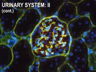
URINARY SYSTEM RENAL CORPUSCLE BOWMAN’S CAPSULE + GLOMERULUS FILTRATION APPARATUS OF KIDNEY 1. BOWMAN’S CAPSULE: - the beginning of the nephron that consists of a blind sac lined with simple squamous epithelium that is continuous with the PCT - parietal layer & visceral layer (specialized) 2. GLOMERULUS: - specialized tuft of capillaries which housed in the capsular space (10-20 capillary loops) - blood flowing through glomerulus capillaries undergoes a filtration process to produce the initial urine filtrate
URINARY SYSTEM RENAL CORPUSCLE BOWMAN’S CAPSULE + GLOMERULUS FILTRATION APPARATUS OF KIDNEY VASCULAR POLE URINARY POLE GLOMERULUS (FILTRATION MEMBRANE): 1- fenestrated capillaries; discontinuous endothelium; fenestrae have a diameter of 500-1000Å and lack a diaphragm 2- continuous basal lamina 3- podocytes of visceral layer; processes contact basal lamina and are separated by slits measuring approximately 250Å
MR CL capsule CORTEX MEDULLA MR AV CL MR KIDNEY H&E
KIDNEY H&E GLOMERULUS MEDULLARY RAY CORTICAL LABYRINTH MEDULLARY RAY GLOMERULUS
RC RC RC RC RC RC RC RC KIDNEY H&E MEDULLARY RAY MEDULLARY RAY MEDULLARY RAY CORTICAL LABYRINTH MEDULLARY RAY
URINARY SYSTEM CORTEX: CORTICAL LABYRINTH 1- RENAL CORPUSCLES 2- PROXIMAL CONVOLUTED TUBULES - longer than DCT and thus more numerous - stain slightly darker and have a larger diameter than DCT - cells are larger and have an irregular luminal surface due to the presence of a “brush border” and glycocalyx 3- DISTAL CONVOLUTED TUBULES - shorter than PCT and thus less prevalent - stain slightly lighter and have a smaller diameter than PCT - cells are smaller and cuboidal, thus more nuclei are apparent in a cross section of a DCT - luminal surface is more uniform since it lacks a brush border
CORTEX: CORTICAL LABYRINTH Bowman’s capsule 1- RENAL CORPUSCLES 2- PROXIMAL CONVOLUTED TUBULES 3- DISTAL CONVOLUTED TUBULES glomerulus RENAL CORPUSCLE DCT PCT KIDNEY H&E
URINARY SYSTEM CORTEX: CORTICAL LABYRINTH 1- RENAL CORPUSCLES 2- PROXIMAL CONVOLUTED TUBULES - 80-90% of H2O and NaCl in glomerular filtrate is reabsorbed in addition to most organic materials: (glucose, proteins, amino acids, etc.) 3- DISTAL CONVOLUTED TUBULES - further resorption of H2O in presence of ADH - sodium resorption in response to aldosterone - calcium resorption in response to PTH
DCT VASCULAR POLE PCT glomerulus URINARY POLE KIDNEY SILVER METHENAMINE
DCT PCT DCT KIDNEY SILVER METHENAMINE
DCT PCT PCT PCT PCT DCT PCT KIDNEY H&E
PCT DCT glomerulus DCT DCT PCT PCT PCT PCT KIDNEY H&E
DCT EA AA DCT DCT glomerulus peritubular capillary plexus PCT PCT KIDNEY H&E
1- STRAIGHT PORTIONS OF PROXIMAL TUBULE (THICK DESCENDING) MEDULLARY RAY 2- STRAIGHT PORTIONS OF DISTAL TUBULE (THICK ASCENDING) 3- COLLECTING DUCTS KIDNEY SILVER METHENAMINE
URINARY SYSTEM KIDNEY H&E CORTEX: MEDULLARY RAY 1- STRAIGHT PORTIONS OF PROXIMAL TUBULE (THICK DESCENDING) 2- STRAIGHT PORTIONS OF DISTAL TUBULE (THICK ASCENDING) TD CD 3- COLLECTING DUCTS - cells are cuboidal in cortex and become progressively more columnar in the medulla and papilla CD - cells stain very lightly with well-defined boundaries - transport urine from nephron to excretory ducts and aids in further H2O resorption in the presence of ADH
KIDNEY H&E COLUMNAR COLLECTING DUCTS NEAR RENAL PAPILLA
URINARY SYSTEM THE NEPHRON & COLLECTING DUCTS MEDULLA: OUTER ZONE 1- STRAIGHT PORTIONS OF PROXIMAL TUBULE (THICK DESCENDING) 2- STRAIGHT PORTIONS OF DISTAL TUBULE (THICK ASCENDING) 3- THIN SEGMENTS OF LOOP OF HENLE (DESCENDING & ASCENDING) 4- COLLECTING DUCTS INNER ZONE 1- THIN SEGMENTS OF LOOP OF HENLE (DESCENDING & ASCENDING) 2- COLLECTING DUCTS
KIDNEY H&E OUTER MEDULLA CD TA CD TD TL
KIDNEY H&E OUTER MEDULLA VASA RECTA TA TL TL TA
KIDNEY H&E INNER MEDULLA TL CD CD TL CD CD TL
ALDOSTERONE SECRETION ANGIOTENSINGOGEN (PLASMA PROTEIN) RENIN ANGIOTENSIN I ANGIOTENSIN II VASOCONSTR URINARY SYSTEM JUXTAGLOMERULAR APPARATUS MACULA DENSA + JUXTAGLOMERULAR (JG) CELLS REGULATE BLOOD FLOW THROUGH GLOMERULUS MACULA DENSA - cells located in the DCT in close contact with the glomerulus and the afferent and efferent arterioles JG CELLS - specialized smooth muscle cells in the wall of the afferent arteriole which contain and secrete RENIN to regulate blood flow through the glomerulus
ALDOSTERONE SECRETION ANGIOTENSINGOGEN (PLASMA PROTEIN) RENIN ANGIOTENSIN I ANGIOTENSIN II VASOCONSTR URINARY SYSTEM JUXTAGLOMERULAR APPARATUS MACULA DENSA + JUXTAGLOMERULAR (JG) CELLS REGULATE BLOOD FLOW THROUGH GLOMERULUS BARORECEPTOR THEORY - assumes JG cells function as stretch receptors (high blood pressure would inhibit release of renin) MACULA DENSA THEORY - assumes the secretion of renin is regulated by the composition of the fluid in the DCT / afferent arteriole (low sodium would increase the release of renin)
ALDOSTERONE SECRETION ANGIOTENSINGOGEN (PLASMA PROTEIN) RENIN ANGIOTENSIN I ANGIOTENSIN II VASOCONSTR URINARY SYSTEM JUXTAGLOMERULAR APPARATUS MACULA DENSA + JUXTAGLOMERULAR (JG) CELLS REGULATE BLOOD FLOW THROUGH GLOMERULUS
KIDNEY H&E MACULA DENSA
KIDNEY H&E RENIN GRANULES JG CELLS AA
1- mucosa lined with transitional epithelium 2- usually lacking submucosa 3- muscularis best developed in ureters (2-3 layers) and bladder (3 layers) URINARY SYSTEM KIDNEY CALYCES/ URETER BLADDER URETHRA
URETER H&E 1: IL 2: OC MUCOSA LAMINA PROPRIA MUSCULARIS ADV
URINARY SYSTEM URETER
URINARY SYSTEM URETER
URINARY SYSTEM BLADDER
URINARY SYSTEM BLADDER transitional epithelium