Learning and Memory
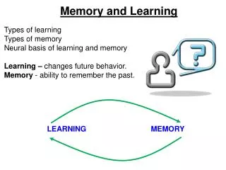
Learning and Memory. Learning. Set of processes by which experience changes the nervous system, changing behavior Resulting changes are memories Nondeclarative memories Declarative memories Enduring changes to the neural circuits. Mechanisms of learning – Synaptic plasticity.

Learning and Memory
E N D
Presentation Transcript
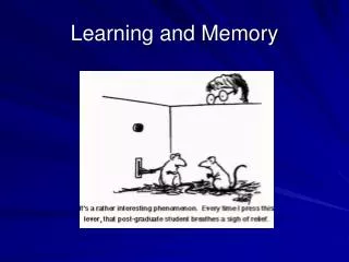
Learning • Set of processes by which experience changes the nervous system, changing behavior • Resulting changes are memories • Nondeclarative memories • Declarative memories • Enduring changes to the neural circuits
Mechanisms of learning – Synaptic plasticity • Synaptic plasticity • Changes in synaptic structure and biochemistry • Long-term potentiation (LTP) • Change in the strength of synaptic connections • Results from repeated activation
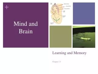
Hippocampal formation - anatomy • Part of the limbic system, located in the temporal lobes • Composed of: • Dentate gyrus, CA1-3 & subiculum • Perforant pathway • Entorhinal cortex to dentate gyrus • Primary source of input
Experimental induction of LTP • Stimulating electrode inserted into the perforant pathway, recording electrode inserted into the dentate gyrus • Single burst of stimulation delivered to the perforant pathway • Resulting EPSP recorded in the neuron population in the dentate gyrus • Provides a baseline measure of normal synaptic firing strength
Experimental induction of LTP • To induce LTP – rapid burst of electrical pulses is delivered to the perforant pathway (~100 pulses/2 seconds) • To detect the presence of LTP - a single, short stimulating burst delivered to the perforant pathway, the population EPSP is measured in the dentate gyrus • Increased response in the dentate gyrus = LTP has occurred • Synapses have been strengthened
LTP characteristics • Synaptic transmission more likely to cause an action potential in the post-synaptic neuron • Lasts from several minutes to years • Can be induced throughout the brain
Associative long-term potentiation • Hebbian rule (Donald Hebb): “Neurons that fire together, wire together” • Synapses that are reliably active just before generation of an action potential are strengthened • Simultaneous firing at a weak and a strong synapse on the same post-synaptic neuron strengthens the weak synapse by association • THIS is how associations are learned! • Ex. Learning to type
Receptor involvement in LTP • Synaptic strengthening depends on: 1. Neurotransmitter binding at the synapse 2. Simultaneous depolarization of the post-synaptic cell • Depolarization of a neuron does NOT strengthen ALL synapses… only those that are active at the time of depolarization
NMDA receptors and LTP • LTP relies on calcium influx at NMDA glutamate receptors • Calcium channels controlled by the NMDA receptor are blocked by a magnesium ion • Magnesium ion is ejected by: 1. simultaneous glutamate binding AND 2. depolarization of the post-synaptic cell (by activity at AMPA receptors on the membrane)
Dendritic spike – an action potential results in a backwash of depolarization up the cell body and dendrites Dendritic spike + glutamate binding at NMDA receptor = calcium channels open to allow calcium influx Strengthening synapses
Role of calcium in LTP • Calcium is critical to establishing LTP • Second messenger activates protein kinases, which influence chemical reactions in the cell necessary for LTP
Strengthening synapses • Three synaptic modifications will support LTP • Addition of receptors • Addition of synapses • Increased glutamate release from the presynaptic membrane
Synaptic modifications supporting LTP – Increased receptors • Individual synapses are strengthened by an increase in AMPA receptors on the post-synaptic membrane • Increases the cell’s response to glutamate release • Hypothesized mechanism: • Calcium activates the CaMK enzyme • Activated CaMK binds to an intracellular portion of the NMDA receptor • Linking proteins bind to the CaMK • AMPA receptors bind to the linking proteins and are embedded into the cell membrane
Synaptic modifications supporting LTP – Synaptogenesis • LTP results in the multiplication of synapses • Most synapses are located on dendritic spines • LTP results in division and multiplication of these spines • Mechanism: • Postsynaptic density expands until it perforates – splits into multiple densities • Following perforation, the presynaptic active zone splits into corresponding regions • Perforated synapse further divides, until the spine branches • Branched spine ultimately becomes two spines, each containing a synaptic region
Synaptic modifications supporting LTP – Synaptogenesis • Results in the terminal button of one presynaptic neuron synapsing with multiple spines on the postsynaptic neuron • Increases communication potential between the two cells • Threefold increase in synapses has been found experimentally
Synaptic modifications supporting LTP – Presynaptic changes • LTP is associated with an increase in glutamate release by the presynaptic neuron • Influenced by retrograde messengers • Nitric oxide – major retrograde signal from NMDA receptors to the presynaptic membrane • NO is synthesized in the postsynaptic membrane in response to calcium influx • Unstable and short-lived, can only diffuse across the synapse before breaking down • Acts as a limited, direct messenger
Long-term depression • Opposite of LTP, long-term depression is a long-lasting weakening of synapses that are not associated with strong inputs/production of action potentials • Seen when two inputs are stimulated at significantly different times, or when a synapse is activated while a cell is weakly depolarized or hyperpolarized • Results in the removal of AMPA receptors from the synapse • Weakening of synaptic strength may be necessary when new learning eliminates the need for previously established synaptic modifications • Ex. Remembering a new locker combination
Classifications of memory • Declarative memory - explicit and readily available to conscious recollection • Episodic – memories of events • Semantic memories – memories of facts • Nondeclarative memory - implicit, unconscious knowledge • Perceptual – memory of previously experienced stimuli • Motor (procedural) – learned behavioral sequences • Stimulus-response – learned responses to specific stimuli
Perceptual learning • Neural changes that result in recognizing a stimulus that has been perceived before • Ex. Learning to recognize the face of a new acquaintance • Allows us to identify people, objects & sensations • New stimuli; changes in previously experienced stimuli
Perceptual learning • Based on synaptic changes in the sensory association cortices • Sensory input activates these brain regions; later input from the same stimulus results in the same pattern of activation • Recognition of the stimulus
+ Classical conditioning • Learning a specific behavioral response in the presence of a given stimulus • Response to an association between two stimuli • Simple, automatic responses • Stimulus-response learning
+ Steps in classical conditioning • Neutral stimulus (NS) has no effect on the subject • Unconditioned stimulus (US) elicits an unconditioned response (UR) • NS is paired repeatedly with the US; UR occurs • NS is presented alone, UR occurs • NS is now the conditioned stimulus (CS)
Neural mechanisms of classical conditioning • Conditioned emotional response – common model of classical conditioning • Demonstrated in footshock paradigm (fear conditioning) • Tone + Footshock = Freezing behavior • Emotional conditioning relies on the amygdala • LTP is exhibited in the amygdala following fear conditioning
Neural mechanisms of classical conditioning • Lateral amygdala receives input on both the CS (tone) and US (footshock) • Prior to learning, CS signal forms weak synapses, US signal forms strong synapses • Neurons in lateral amygdala receive these signals, project to the central amygdala (CNA) • CNA – generates emotional response (UR: freezing) • Strong synapses from US reliably produce an action potential in projections to CNA • Synaptic activation at weak CS synapses + depolarization by US signal strengthens CS synapses • CS/US association is formed
Hebb’s rule • Neurons that fire together, wire together • Demonstrated by the strengthening of the connection between neurons signaling the CS and neurons producing the behavioral response • Repeated firing of the weak tone synapse + footshock-produced depolarization strengthens the tone synapse Firing at the tone synapse will now independently produce an action potential resulting in freezing behavior.
Motor learning • Changes that result in a new sequence of movements (Procedural memories) • Establishes new motor skill sequence • Based on changes in the motor system • New behaviors require extensive modification of brain circuits; adjustments produce changes to these circuits • Learning to walk vs. learning to run, skip and dance
Neural control of motor learning • Learning a new sequence of motor response involves sensory input and motor output • Two pathways connect sensory and motor association cortices: • Direct transcortical projections • Connections through thalamus and basal ganglia
Neural control of motor learning • Initial learning of a complex behavior requires intense focus on environmental stimuli and processing of sensory input • Accomplished by transcortical pathways between sensory and motor association cortices • As the complex behavior is repeated, behavior becomes more automatic • Processing is transferred to the basal ganglia
Neural control of motor learning • Basal ganglia receives input from sensory association areas, and prefrontal cortex (planning) • Projects to the prefrontal motor association area, which initiates motor output • Repetition strengthens the synapses between sensory inputs and motor outputs • Cortex becomes less involved • Lesions of the basal ganglia disrupt motor learning and performance of learned motor behaviors
Operant conditioning • Learning to make a response in order to gain reinforcement or avoid punishment • Formation of associations between a discriminative stimulus, behavioral output, and resulting consequences • Discriminative stimulus: contextual cue • In response to the discriminative stimulus, behavior occurs • Reinforcing or punishing stimulus follows the behavior • Animal learns to make the correct behavior in the context, in order to gain reinforcement/avoid punishment
Operant conditioning • Behaviors increase when the consequences are favorable, decrease when outcomes are aversive • Learning from our experiences: figuring out behaviors to repeat, and other behaviors not to repeat • Stimulus-response learning
Reinforcement • Outcomes that increase the likelihood of a behavior • Neural reinforcement mechanisms strengthen synapses between neurons that detect discriminative stimuli and neurons that produce a behavioral response
Neural circuitry of reinforcement • Neural circuitry involved in reinforcement: • Medial forebrain bundle (MFB) – axon bundle that extends from the VTA to the NAc, passing through the lateral hypothalamus • Stimulation of the MFB is highly rewarding • Common model of reward motivation
Neural circuitry of reinforcement • Mesolimbic system – dopamine neurons that project to the amygdala, hippocampus, and nucleus accumbens(NAc) – major system involved in reward motivation • Dopamine release in the NAc is highly reinforcing • Human research supports a role for the NAc in reinforcement: • fMRI: NAc activation when expecting money or sex
Neural circuitry of reinforcement • Detection of reinforcing stimuli involves input from regions that project to the VTA • Amygdala – detects emotionally relevant stimuli • Determines the reinforcing value of stimuli • Lateral hypothalamus – involved in seeking and detecting biologically relevant stimuli • Signals the presence of reinforcing stimuli • Prefrontal cortex – evaluates sensory stimuli, makes strategies and evaluates outcomes • Signals that behavior is succeeding
Strengthening of synapses by reinforcement Dopamine axons from the VTA and glutamate axons from hippocampus, amygdala and prefrontal cortex synapse on the same NAc cells NAc projects to basal ganglia, influencing behavioral output Depolarization of NAc neurons by DA (reinforcement) strengthens the glutamatergic synapses, increasing the likelihood of reinforced behaviors Neural circuitry of reinforcement
Relational learning • Complex learning involving associations between multiple stimuli, contexts, behaviors and outcomes • Most learning involve relational learning • Requires learning of individual stimuli, and how each stimulus is related to the others • Examples: • Episodic learning – establishing memories of experiences • Spatial learning – forming memories of where objects are located in space • Observational learning – social learning in which the behaviors of others are observed and replicated
Hippocampus and relational learning • Hippocampus is critical to relational learning • NMDA receptors in the hippocampus • Lack of NMDA receptors prevents the establishment of LTP in the hippocampus and impairs spatial task learning • Mice with a genetic mutation for more efficient NMDA receptors exhibit greater EPSPs in the hippocampus and learn a spatial task much faster than control mice
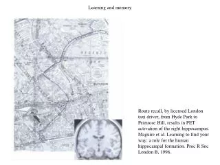
Spatial memory • Memory of the location of objects and places in space • Relies on the right hippocampal formation • Damage to this area produces profound deficits in spatial memory • PET shows increased activity in this region while recalling spatial locations and navigating through an environment • Taxi driver study
Hippocampus and spatial memory • Hippocampus is not necessary for most simple stimulus-response learning; it IS critical for relational learning • Studied in the Morris water maze - measure of spatial learning • Animal model of relational learning
Hippocampus and spatial memory • Animals and humans with hippocampal lesions can learn stimulus-response tasks • Animals with lesions can perform well in the MWM if released from the same spot every time – simple stimulus-response learning • Animals with hippocampal lesions fail to learn spatial relations, and cannot navigate according to contextual cues • Animals with hippocampal lesions fail at the MWM if released from a different location every time
Hippocampal place cells • Place cells - individual cells in the hippocampus that fire only when an animal is in a particular location • Each place cell responds maximally to one location, known as its spatial receptive field
Hippocampal place cells • Place cells respond based on environmental cues about location • Do not intrinsically know where the animal is located • Same arrangement of environmental cues in two different locations identical place cell response • Cues that indicate a difference in environments different place cell response • Place cells aid in spatial learning by providing a signal about where the animal is in space • Place cells are concentrated in the dorsal hippocampus in rats; posterior hippocampus in humans
Human anterograde amnesia • Anterograde amnesia – loss of relational learning ability • New declarative memories are not formed • Simple stimulus-response, perceptual and motor learning abilities remain intact • Previously formed memories remain intact • Retrograde amnesia – loss of previously formed declarative memories
Development of anterograde amnesia • Appearance of anterograde amnesia typically includes some retrograde amnesia • May be loss of hours, days or years • Results from bilateral damage to, or removal of the medial temporal lobes • Unilateral damage may produce minor memory deficits
Development of anterograde amnesia • Famously discovered in H.M. • Both medial temporal lobes were removed to treat severe epilepsy • Resulted in pervasive anterograde amnesia, accompanied by some retrograde amnesia
Development of anterograde amnesia • Korsakoff’s syndrome • Brain damage to the mammillary bodies resulting in anterograde amnesia • Caused by a lack of vitamin B1 (thiamine) in the brain • Typically the result of severe alcoholism
Anatomy of amnesia • Medial temporal lobe contains the hippocampus – critical to memory formation • Input to the hippocampus: from the cingulate cortex and cortical association areas, via entorhinal cortex • Output: back to cingulate cortex and cortical association areas, through entorhinal cortex • Damage to the hippocampus, or its inputs or outputs, results in anterograde amnesia