Antenatal Surveillance
Antenatal Surveillance. Ahmet Baschat, MD Professor Head, Section of Fetal Therapy Dep. of Ob/Gyn, Reprod. Sciences University of Maryland School of Medicine. Antenatal surveillance. AIM: to prevent compromise & stillbirth REQUIREMENTS Know limitations of surveillance tests


Antenatal Surveillance
E N D
Presentation Transcript
Antenatal Surveillance Ahmet Baschat, MD Professor Head, Section of Fetal Therapy Dep. of Ob/Gyn, Reprod. Sciences University of Maryland School of Medicine
Antenatal surveillance • AIM: to prevent compromise & stillbirth • REQUIREMENTS • Know limitations of surveillance tests • Recognize specific maternal risk factors • Understand progression of maternal disease • Understand progression of fetal disease • Physical evaluation of the fetus • PRINIPAL DECISIONS: • Is delivery indicated ? • Are steroids indicated ? • When should the patient be seen again ?
Two important principles • The need for intervention is based on the balance of fetal risks versus neonatal risks • The monitoring interval has to be based on the speed of clinical progression
adaptation Fetal condition acidemia compromise Stillbirth Pathways of deterioration Not every condition produces the same clinical findings with fetal compromise… INTERVENTION hypoxemia • alterations fetal heart rate pattern • declining amniotic fluid volume • decrease in dynamic variables • alterations in regional blood flow
Surveillance tests • Maternal history and risk factors • Fetal physical examination • Anatomy • Size • Proportion • Growth • Amniotic fluid volume • Biophysical variables • Heart rate parameters • Cardiovascular parameters
Maternal risk factors • Current pregnancy • specific referral • Hypertension • Pre-eclampsia • Gestational Diabetes • Prior pregnancy • Pre-eclampsia • Stillbirth / Losses • Abruption • Medical Illness • Hypertension • Diabetes • Lupus • Thrombophilia • Recognition of maternal risk factors is essential because it determines which tests should be performed and at which frequency. • A thorough history and physical examination should form part of the initial assessment of the patient. • Additional laboratory studies may be indicated to clarify diagnoses and prognoses.
Fetal risk factors • Chromosomal abnormalities, fetal syndromes and viral infections mimic many potentially treatable fetal conditions. • Detailed anatomic survey is therefore essential • Features of aneuploidy • Multiple malformations • Multiple markers • Abnormal growth • Features of Syndromes • Recognized combinations of physical abnormalities • Viral infection • Echogenicities in organs • Fluid accumulation in body cavities • Abnormal growth • These differential diagnoses must be considered at each visit.
Fetal size • BPD • HC • TCD • AC • FL • HL • SEFW • Fetal size is measured by • Head size • Biparietal diameter (BPD) • Head circumference (HC) • Cerebellar diameter (TCD) • Body size • Abdominal circumference (AC) • Skeletal size • Femur length (FL) • Humerus length (HL) • Estimated fetal weight (EFW) • Composite varieble • Assessment of size requires reference ranges and knowledge of gestational age.
Percentile Percentile Percentile 25 25 25 5 5 5 20 20 20 40 40 40 Gestational age Gestational age Gestational age Symetrical small Asymmetrical – short bones Asymmetrical – small abdomen Fetal proportion • Measurements of fetal symmetry: • Head to abdomen ratios • (HC/AC) • (TCD/AC) • Head to Femur ratio (BPD/FL) • Femur to abdomen ratio (FL/AC) • Asymmetrically abnormal size: • Early growth delay • Skeletal dysplasia • Trisomy • Syndromes • Symmetrically abnormal size • Severe growth delay • Aneuploidy • Viral infection
AC AC AC AC AC AC AC AC AC AC AC AC Fetal growth Percentile • Growth is dynamic: single and serial measurements at >14 day intervals are needed. • Continued growth along reference ranges is most likely normal. • measurements that fall off the curve are likely abnormal. • Abnormal head growth can indicate aneuploidy or viral infection • Abdominal circumference: single best measure of fetal nutrient status. • Skeletal growth abnormality: important marker for skeletal dysplasia. = normal = abnormal 25 5 20 40 Gestational age
Amniotic fluid volume • Amniotic fluid index • Sum of 4 quadrant vertical pockets • Allows trend-analysis • Subjectively reduced fluid • Maximum pocket < 3 cm • No fetal bladder filling • Empty fetal stomach • Restricted fetal movement • Flexed fetal position • Uterine molding around fetus • Deceleration with movement • Deceleration with transducer pressure • increased uterine contractility • Single vertical pocket < 2cm After 14 wks a measure of - fetal urine production - placental fluid exchange
Amniotic fluid volume • Fluid volume is determined by the relative rate of production (urination) and removal (fetal intake). • If conditions co-exist dynamics may appear normal (i.e. placental insufficiency and maternal diabetes. • ↑ fluid – polyhydramnios • Maternal diabetes • Tracho-esophageal fistula • Choanal atresia • Aneuploidy • Viral infection • Tachycardia • Twin-twin transfusion • ↓ fluid – oligohydramnios • Rupture of membranes • Placental insufficiency • Viral infection • Aneuploidy • Urinary obstruction • Twin-twin transfusion
1 2 3 4 5 Doppler ultrasound • This standardized approach to the Doppler examination of every vessel is essential in order to achieve reproducible and reliable results: • Zoom to the area of interest • Apply color Doppler • Narrow color box • Adjust velocity scale • Apply pulsed wave gate • Adjust gate to cover vessel • Adjust velocity scale • Adjust filter • 3-5 uniform waveforms • No fetal activity
Doppler ultrasound Velocity • Continuous trace of the waveform from start to the beginning of the next • In venous vessels automatic tracing software should not be used because the triphasic waveform is not appropriately analyzed Pulsatility index S systolic peak velocity (S - D) TAMX D end-diastolic peak velocity TAMX Time • The Pulsatility index is recommended for arterial vessels • The Pulsatility index for veins is recommended for venous vessels • Reference ranges should be used to interpret the Doppler values Pulsatility index systolic peak velocity S (S - a) D diastolic peak velocity TAMX TAMX atrial systolic peak velocity a
S D Vessel tone Vascular histology Arterial Doppler • Relationship of systole and diastolic velocity and waveform characteristics depend on • Input pressure • Vascular resistance • Vascular resistance may be altered due to • Changes in vessel tone • Structural vascular change Input pressure Peripheral resistance • Autoregulation • MCA • Renal • Hepatic • Adrenal • Coronaries • Splenic • Failed placentation • Umbilical arteries • Uterine arteries • Vasoconstriction • DA after • Indomethacin
S D A Afterload 60 - 70% Placenta Venous Doppler • Venous Doppler gives information about forward cardiac function • Compliance • Relaxation • Contractility • Afterload • All vessels have the same waveform • Systolic peak • Diastolic peak • Atrial systole • Clinically most studied • Ductus venosus • Inferior vena cava • Umbilical vein Compliance Contractility
Maternal compartment 500-600 ml/min. 11 weeks 24 weeks 40 weeks 12 m2 40 weeks 24 weeks 11 weeks 250 ml/Kg/min. Fetal compartment The placenta • A two compartment nutrient, fluid and gas exchange organ • Maternal compartment • Uterine artery Doppler. • Fetal compartment • Umbilical artery Doppler. • Maturation of the vasculature is observed in both compartments, • Loss of uterine artery notch • Appearance of umbilical diastolic velocity • Successive decline in Pulsatility index in both vessels • Gestational age is important for assessing waveforms trophoblast invasion villous branching
The placenta Uterine artery Umbilical artery • Abnormal trophoblast invasion: • High uterine artery PI • Persistent uterine artery notch • Abnormal villous vascular tree • Umbilical artery Doppler. • Fetal compartment • 30% abnormal villous vasculature – high umbilical artery PI. • 50% abnormal villous vasculature – absent umbilical artery end-diastolic velocity • 70% abnormal villous vasculature – reversed umbilical artery end-diastolic velocity • Risk for hypoxemia / acidemia proportional to decrease in umbilical end-diastolic flow
Middle cerebral artery • Branch of the circle of Willis • Use parietal bone window • Parallel to wings of sphenoid • Proximal part recommended • Insonate at 0 degrees • Two parameters are of importance in this vessel • Decreased pulsatility index in • Fetal hypoxemia • Fetal hypertension • Both are indistinguishable by the waveform. • Increased peak systolic velocity (0 degree insonation) in • Fetal anemia • Increased paCO2
Ductus venosus • Is the primary shunt regulating nutrient flow to the liver and heart • Can be imaged in a saggital or abdominal transverse view. • From the first trimester on the a-wave should be antegrade • Pulsatility index for veins significantly decreases with advancing gestation.
Umbilical vein • Examine in the straight abdominal portion or cord • 90% of fetuses have constant flow from 12 weeks on. • Pulsations can be • Monophasic • Biphasic • Triphasic • Monophasic pulsations are relevant if central veins are abnormal • Multiphasic pulsations indicate abnormally high venous pressure • Clinical applications: • Fetal growth restriction • Twin-twin transfusion • Hydrops
Umbilical vein Abnormal veins • The following are abnormal • Decreased a-wave • Decreased D-wave • Decreased v-trough • These abnormalities produce an increase in the Pulsatility index for veins • Absent or reversed flow during the a-wave gives a simple visual assessment of abnormal ductus venosus flow Constant
Stable constellation of activity Rest activity cycles Vibroacoustic Glucose & breathing Movement & FHR Breathing movement Gross body movement Fetal behavior 1st trimester 2nd trimester 3rd trimester Behavioral states Cyclicity Coupling • Fetal behavior develops sequentially: • Isolated activity • Coupling of behavior • Rest activity cycles • Behavioral states • Movement frequency is determined by gestational age and behavioral state Activity
Fetal tone & movement • Fetal tone can be assessed by examining flexion-extension of the extremities and/or the trunk. • Absence can be explained by • Fetal hypoxemia • Fetal acidemia • Fetal rest • Neuromuscular block • CNS abnormality • Best interpreted in the context of a full biophysical profile score
Fetal breathing • Chest movement, diaphragm movement and hiccups count • Absence can be explained by • Fasting state • Fetal hypoxemia • Fetal acidemia • Fetal rest • Neuromuscular block • CNS abnormality • Absence of fetal breathing should prompt re-evaluation after maternal food intake.
cerebral cortex VMC RAS ANS CVS Heart BP = CO peripheral resistance x stroke volume HR x Fetal heart rate • A record of autonomic regulation of intrinsic cardiac activity and its modulation by regulatory centers. • Vasomotor center (VMC) • Reticular activating system (RAS) • Autonomic nervous system (ANS) • Analyzed visually by • Baseline heart rate • Reactivity • Variability • Periodic changes • Computerized analysis • Short term variation (ms)
Fetal heart rate • Reactivity virtually excludes hypoxemia • Causes of non-reactivity • Gestational age • Behavioral state • Hypoxemia / Acidemia • Medications • Variable decelerations • Cord compression • Late decelerations • >8 torr drop in paO2 • Hypoxemia • Short term variation <3.5 ms • Hypoxemia • Abnormal brain development
Biophysical profile score For each component presence = 2 points, absence = 0 points • Modified BPS • Amniotic fluid index • Reactive FHR • Composite score of 5 variables • Normal = 10, 8 (PNM=1/1000) • Equivocal (PNM=7-10/1000) • 8 with oligohydramnios • 6 • Abnormal (PNM=12-300/1000) • 6 with oligohydramnios • 4,2,0 • Score of 4 – immediate retesting for 30 min • Persistent score of 4, or less – immediate delivery Tone at least one episode of active limb, trunk or hand extension with return to flexion Movement at least 3 discrete body/limb movements (active continuous considered as single movement) at least one episode of at least 30 seconds duration (includes hiccups) Breathing Amniotic fluid at least one single vertical pocket >2 cm Heart rate • at least 2 acceleration of • - 10 beat x 10 sec (24-28 weeks) • 15 beat x 15 sec (28-34 weeks) • 20 beats x 20 sec (>34 weeks) Manning et al., Am J Obstet Gynecol 1982
-reaktiv pH < 7.20 pH < 7.10 FHR parameters biophysicalparameters DopplerParameter 0 LTV <30 STV <3.5 Tone & Movement Breathing -2 Δ pH -4 -6 -8 abnormal cCTG und Ductus venosus comparable pH biophysical parameter = closer relationship with pH -10 NST cCTG* AFV FBM FGM Tone AEDV TAO DAO MCA CPR DV Akalin-Sel et al., Arduini et al., Bilardo et al., Guzman et al,, Hecher et al,, Nicolaides et al., Ribbert et al., Rizzo et al., Soothill et al., Visser et al., Weiner et al.
Summary Tests provide specific information Fetal anatomy – differential diagnosis Fetal growth – placental performance Amniotic fluid – volume status / placental transfer Uterine Doppler – trophoblast invasion Umbilical Doppler – vascular exchange area MCA Doppler – pCO2, Hgb, Oxygen, Hypertension Venous Doppler – rhythm, forward cardiac function Dynamic variables – Maturation, Behavioral state, pO2 FHR variables – CNS, PNS, pO2 Specific conditions require specific tests…
adaptation Fetal condition acidemia compromise Stillbirth Pathways of deterioration Not every condition produces the same clinical findings with fetal compromise… INTERVENTION hypoxemia • alterations fetal heart rate pattern • declining amniotic fluid volume • decrease in dynamic variables • alterations in regional blood flow
Integrated fetal testing • Every surveillance test has advantages and disadvantages • Integrated fetal testing combines different tests as needed • Distinguishing false positives from true positives • Detect different avenues of fetal deterioration • Examples of integrated testing • Biophysical profile score • Fetal Apgar Score • Integrated fetal testing management
* * * * Middle cerebral artery Increased baseline Decreased variation / variability Decreased reactivity variation decrease / loss BPS Delayed maturation of FHR control declining amniotic fluid volume declining global activity Loss of breathing Delayed behavioral maturation 0 Δ pH - 2 - 4 - 6 CIRCULATORY COMPROMISE Ductus venosus CIRCULATORY DECOMPENSATION PLACENTA – BASED GROWTH DELAY Umbilical artery CIRCULATORY COMPENSATION ABNORMAL BPS DEVELOPMENTAL DELAY DECLINING ACTIVITY Loss of movement Loss of tone STILLBIRTH HYPOXEMIA ACIDEMIA Baschat 2008
0 Δ pH - 2 - 4 - 6 Ductus venosus PLACENTA – BASED GROWTH DELAY Umbilical artery Middle cerebral artery Nonreactive heart rate declining amniotic fluid volume Loss of breathing ? STILLBIRTH Baschat 2008
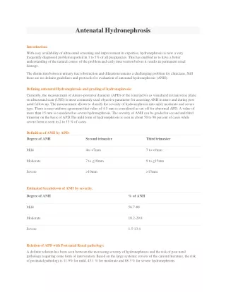
abnormal increased elevated indexAbsent / reversed end-diastolic velocity decreased index decreased ratio Approach to the fetus with small biometry Likely diagnosis Anatomy Aneuploidy Syndrome Viral infection normal Amniotic fluid normal or decreased Umbilical artery Doppler normal IUGR due to placental insufficiency Middle cerebral artery Doppler normal Cerebroplacental ratio normal Constitutionally small fetus normal repeat examination at 14 days
The principal decisions New onset brain sparing Oligohydramnios UA – AEDV / REDV Abnormal DV Doppler Early gestation = high threshold Late gestation = low threshold The monitoring interval Early stages require less frequent monitoring Disease acceleration = ↑ monitoring frequency Which thresholds to base delivery on ?
↑ UA PI ↓ Cerebroplacental Doppler ratio UA A-REDV Brain sparing Abnormal DV index ↑ UA PI DV RAV – UV pulsations ↓ Cerebroplacental Doppler ratio Brain sparing UA A-REDV Abnormal DV index DV RAV – UV pulsations ↓ CPR ↑ UA PI Severe Progressive 27 day latency 30 weeks delivery Degree of Doppler Abnormality 38 day latency / 33.4 weeks delivery Mild 46 day latency / 35.3 weeks delivery 27 29 31 33 35 37 39 Gestational weeks Turan OM et al., Ultrasound Obstet Gynecol 2008
After diagnosis of FGR: • Weekly UA Doppler • Severe deteriorates within 2 weeks • Progressive deteriorates over next 2 weeks • If no change over 4 weeks – probably mild Turan OM et al., Ultrasound Obstet Gynecol 2008
1%/day in utero 100 90 80 2% / day in utero 70 60 50 Percent 40 30 20 10 0 24 25 26 27 28 29 30 31 32 Gestational week N=642 Overall mortality = 130 (21%) Intact survival = 352 (54%) Neonatal survival Intact survival Baschat et al., Obstet Gynecol 2007
2.5 prospective stillbirth rate 2 prospective perinatal mortality rate 1.5 Risk / 1000 ongoing pregnancies 1 0.5 0 24 25 26 27 28 29 30 31 32 33 34 35 36 37 38 39 40 41 42 43 Prospective Stillbirth rate • If Fetal growth restriction is observed at 38 weeks the statistical benefit of delivery outweighs the risk of continuing pregnancy Favor delivery for singletons Kahn et al., Obstet Gynecol, 2002
Divon et al., 1989, AJOG • Clinical trial • IUGR fetuses with A/REDF • Daily BPS • Delivery for • BPS of 4 or less • Oligohydramnios • maternal status • documented lung maturity • No stillbirths, no acidemia at birth
Cosmi et al., 2005, Obstet. Gynecol • 145 idiopathic IUGR; delivery for BPS or CTG • Two groups of fetuses • complete deterioration of all Doppler parameters • Abnormal BPS / CTG with maintained Dopplers • No differences in perinatal outcome • Predictors of outcome • UA REDV • DV REDV • Birthweight • Even with DV A/REDV up to 8 days normal BPP !
100 80 60 Percent ongoing pregnancies 40 20 0 24 25 26 27 28 29 30 31 32 Gestational week Combined tests – hypothetical modeling modified BPP & AREDV DV RAV or UV Pulsations and absent movement or fluid 11/17 stillbirths prevented 12/ 24 Acidemia prevented28.0 weeks delivery GA 18/29 stillbirths prevented17/30 Acidemia prevented29.3 weeks delivery GA 15% increase in survival Abnormal BPP alone 19/29 stillbirths prevented18/30 Acidemia prevented 28.5 weeks delivery GA 8% increase in survival Baschat et al., AJOG 2007
TRUFFLE STV < 3.5 msec STV < 4 msec Steroids beneficial Periviability individualize DV - RAV DV PI > 3SD Greatest survival benefit / day in utero Not well delineated Intervention triggers UA - REDV UA-AEDV Biophysical profile score < 6 Integrated fetal testing score < 8 24 26 29 32 34 Baschat 2008
140 Increased insulin resistance 130 120 110 Higher postprandial glucose 100 90 80 Lower fasting glucose 70 60 Non-pregnant 250 Pregnant 200 150 100 50 0 2 4 6 8 22 24 12 20 10 14 16 18 8 Maternal Diabetes GLUCOSE mg/dl INSULIN Potential risk to develop diabetes in pregnancy IU / ml Risks of worsening glycemic control in existing diabetes
> Age 20 Age 10-19 Age<10 <10 years 10-19 years >20 years <105 mg% >105 mg% <140 mg% >140 mg% Diet Insulin Insulin Benign Retinopathy Nephro-pathy Proliferative Retinopathy Cardiac A 1 A 2 B C D F R H Macrosomia IUGR Diagnosis Current pregnancy Duration Fasting sugar 1’ sugar Therapy Vascular risks Whites Class Fetal death Pregnancy risks Anomalies PIH / PET Maternal Mortality
Once / twice weekly Once / twice weekly Surveillance in diabetes • Signs of glycemia-mediated risks • Macrosomia • Polyhydramnios • Myocardial thickening • Signs of vascular-mediated risks • IUGR • Abnormal uterine artery Dopplers Monitor fluid & FHR Monitor like IUGR Empiric monitoring based on GA Start monitoring in the presence of above signs