Understanding Electronic Absorption Spectroscopy of Aromatic Molecules
490 likes | 604 Vues
Explore how p-conjugation in linear polyenes and aromatic hydrocarbons affects electronic spectra shift and energy gap narrowing. Learn about modeling p-electrons as particles in a box and the absorption spectra of various compounds.

Understanding Electronic Absorption Spectroscopy of Aromatic Molecules
E N D
Presentation Transcript
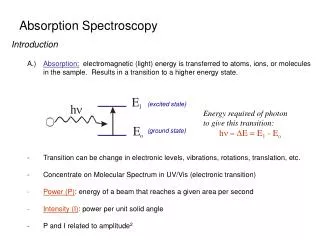
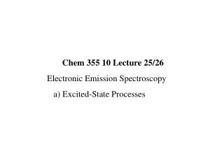
Chem 355 10 Lecture 23 Electronic Absorption Spectroscopy b)Free Electron Model c) Exciton Interactions
The Free Electron Model and Linearpolyene and Aromatic Hydrocarbon Spectra
The Free Electron Model and Linearpolyene and Aromatic Hydrocarbon Spectra
The Free Electron Model and Linearpolyene and Aromatic Hydrocarbon Spectra Porphin Protoporphyrin IX
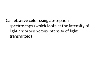
As the p-conjugation in both linear polyenes and aromatic molecules becomes more extended, the electronic spectra shift to longer wavelengths, or as the energy gap between the ground and 1st excited state narrows. While ethylene absorbs deep in the uv (~ 180 nm), b-carotene absorbs far into the visible region producing the orange-yellow colour of carrots, and the even more conjugated lycopenes result in red colour of tomatoes. The p electrons in these types of molecules can be modeled as “particles in a box”, or “ – on a hoop”.
As the p-conjugation in both linear polyenes and aromatic molecules becomes more extended, the electronic spectra shift to longer wavelengths, or as the energy gap between the ground and 1st excited state narrows. While ethylene absorbs deep in the uv (< 180 nm), b-carotene absorbs far into the visible region producing the orange-yellow colour of carrots, and the even more conjugated lycopenes result in red colour of tomatoes. The p electrons in these types of molecules can be modeled as “particles in a box”, or “ – on a hoop”.
As the p-conjugation in both linear polyenes and aromatic molecules becomes more extended, the electronic spectra shift to longer wavelengths, or as the energy gap between the ground and 1st excited state narrows. While ethylene absorbs deep in the uv (~ 180 nm), b-carotene absorbs far into the visible region producing the orange-yellow colour of carrots, and the even more conjugated lycopenes result in the red colour of tomatoes. The p electrons in these types of molecules can be modeled as “particles in a box”, or “ – on a hoop”.
The electronic absorption spectra of ethylene and octatetraenc (C8H10). The p-electrons are considered as particles in a 1-dimensional box. The electrons are considered to be at a constant potential over a distance “a” governed by the extent of the delocalization. The energies are given by: where De represents the energy difference between the highest occupied energy level and the lowest (next) unoccupied level.
The electronic absorption spectra of ethylene and octatetraenc (C8H10). The p-electrons are considered as particles in a 1-dimensional box. The electrons are considered to be at a constant potential over a distance “a” governed by the extent of the delocalization. The energies are given by: where De represents the energy difference between the highest occupied energy level and the lowest (next) unoccupied level.
If the highest occupied level is designated with n = N, then n = N+1 represents the unoccupied level to which the electron will be excited: N+1 N
If the highest occupied level is designated with n = N, then n = N+1 represents the unoccupied level to which the electron will be excited: N+1 N
If the highest occupied level is designated with n = N, then n = N+1 represents the unoccupied level to which the electron will be excited: N+1 N
Consider the case of ethylene in which N = 1. The distance over which the p-electrons are free to move is taken as the bond distance plus a distance beyond the centre of the C-atoms. With a bond distance = 1.35Å an additional 2.5Å is added to account for the additional distance, or a = 3.85Å. Both electrons occupy the lowest level or, 2N+1 = 3. = 163 x 10-9m = 163 nm, deep in the UV = 61,349,7 cm-1
Consider the case of ethylene in which N = 1. The distance over which the p-electrons are free to move is taken as the bond distance plus a distance beyond the centre of the C-atoms. With a bond distance = 1.35Å an additional 2.5Å is added to account for the additional distance, or a = 3.85Å. Both electrons occupy the lowest level or, 2N+1 = 3. = 163 x 10-9m = 163 nm, deep in the UV = 61,349,7 cm-1
Consider the case of ethylene in which N = 1. The distance over which the p-electrons are free to move is taken as the bond distance plus a distance beyond the centre of the C-atoms. With a bond distance = 1.35Å an additional 2.5Å is added to account for the additional distance, or a = 3.85Å. Both electrons occupy the lowest level or, 2N+1 = 3. = 163 x 10-9m = 163 nm, deep in the UV = 61,349,7 cm-1
With octatetraene: CH2=CH–CH=CH–CH=CH–CH=CH2 there are 7 average C–C of 1.4Å = 9.8Å or a =9.8Å. To find N: 2 electrons per level, consistent with the Pauli Exclusion Principle are added: 5 N = 4 3 (33,100 cm-1 or 302 nm observed) 2 1
With octatetraene: CH2=CH–CH=CH–CH=CH–CH=CH2 there are 7 average C–C of 1.4Å = 9.8Å or a =9.8Å. To find N: 2 electrons per level, consistent with the Pauli Exclusion Principle are added: 5 N = 4 3 (33,100 cm-1 or 302 nm observed) 2 1
With octatetraene: CH2=CH–CH=CH–CH=CH–CH=CH2 there are 7 average C–C of 1.4Å = 9.8Å or a =9.8Å. To find N: 2 electrons per level, consistent with the Pauli Exclusion Principle are added: 5 N = 4 3 (33,100 cm-1 or 302 nm observed) 2 1
With octatetraene: CH2=CH–CH=CH–CH=CH–CH=CH2 there are 7 average C–C of 1.4Å = 9.8Å or a =9.8Å. To find N: 2 electrons per level, consistent with the Pauli Exclusion Principle are added: 5 N = 4 3 (33,100 cm-1 or 302 nm observed) 2 1
The electronic spectra of aromatic molecules can be treated in a similar fashion with the difference being that the p-electrons are considered to be particles on a hoop rather than in a 1-dimensional box and moving at a constant potential. This is an extension of the de Broglie approach to quantization by fitting standing waves into a circular planar orbit, The allowed energies are given by: In which m in this case can take on values of 0, ±1, ±2, etc. The 2-fold degeneracy for m > 1 arising from the electron orbiting in a clockwise and anticlockwise direction.
±3 ±2 ±1 0
±3 ±2 ±1 0 For benzene m = M = 1 (highest occupied orbital), r = 1.45Å
±3 ±2 ±1 0 For benzene m = M = 1 (highest occupied orbital), r = 1.45Å
The absorption is in the uv as observed. However, the transition depicted in the previous slide is forbidden by symmetry but becomes weakly allowed (observed) due to an appropriate vibration. The free electron model works reasonably well for molecules with more extensive conjugation and does indicate the general trends of lower-energy transitions associated with increasing electron delocalization. This trend is very apparent in quantum dots (CdSe particles) in which the fluorescence shifts to longer wavelenths with increasing size of the particles (Chem 393 Lab).
The absorption is in the uv as observed. However, the transition depicted in the previous slide is forbidden by symmetry but becomes weakly allowed (observed) due to an appropriate vibration. The free electron model works reasonably well for molecules with more extensive conjugation and does indicate the general trends of lower-energy transitions associated with increasing electron delocalization. This trend is very apparent in quantum dots (CdSe particles) in which the fluorescence shifts to longer wavelenths with increasing size of the particles (Chem 393 Lab).
The absorption is in the uv as observed. However, the transition depicted in the previous slide is forbidden by symmetry but becomes weakly allowed (observed) due to an appropriate vibration. The free electron model works reasonably well for molecules with more extensive conjugation and does indicate the general trends of lower-energy transitions associated with increasing electron delocalization. This trend is apparent in quantum dots (CdSe particles) in which the absorption and fluorescence shift to longer wavelenths with increasing size of the particles (Chem 393 Lab).
The delocalization found in quantum dots involves the exchange of electrons between atoms in the crystalline state and is more apply referred to in terms of excitonic states. The formation of excitons will be considered next.
Similarly, coulombic and exchange interactions occur between electronically-excited, and ground-state molecules.
Similarly, coulombic and exchange interactions occur between electronically-excited, and ground-state molecules. The coulombic interactions occur over a distance and can be approximated in terms of dipole-dipole interactions.
Similarly, coulombic and exchange interactions occur between electronically-excited, and ground-state molecules. The coulombic interactions occur over a distance and can be approximated in terms of dipole-dipole interactions. Exchange interactions require orbital overlap between the interacting partners and are therefore short-range in nature.
Similarly, coulombic and exchange interactions occur between electronically-excited, and ground-state molecules. The coulombic interactions occur over a distance and can be approximated in terms of dipole-dipole interactions. Exchange interactions require orbital overlap between the interacting partners and are therefore short-range in nature. The oscillating transition dipoles that characterize the coulombic interactions are associated with a characteristic distance and orientation dependence
In spin resonance spectroscopy dipoles aligned by the magnetic field interact with a parallel or anti-parallel geometry. a = q cos q b = q cos qg = 0 cosg = 1
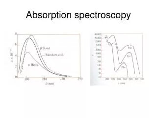
Exciton splitting. i) Exciton interactions split the absorption spectrum and re- . distribute the intensity into the resulting bands. To a first . approximation the total intensity remains unchanged. ii) A significant biophysical example of this effect is the . alterations that occur in the uv absorption band of . polypeptides when they adopt a-helical, b-sheet or . random coil conformations.
Exciton splitting. i) Exciton interactions split the absorption spectrum and re- . distribute the intensity into the resulting bands. To a first . approximation the total intensity remains unchanged. ii) A significant biophysical example of this effect is the . alterations that occur in the uv absorption band of . polypeptides when they adopt a-helical, b-sheet or . random coil conformations. iii) The following CD spectra provides a demonstration of . weak coupling between 2 molecules resulting in an . exciton splitting
Exciton splitting. i) Exciton interactions split the absorption spectrum and re- . distribute the intensity into the resulting bands. To a first . approximation the total intensity remains unchanged. ii) A significant biophysical example of this effect is the . alterations that occur in the uv absorption band of . polypeptides when they adopt a-helical, b-sheet or . random coil conformations. iii) The following CD spectra provides a demonstration of . weak coupling between 2 molecules resulting in an . exciton splitting
Adenosine displays a weak CD spectra due to the asymmetric perturbation of the ribose moiety on the planar adenine moiety. (c)
polyA (a) and the even the dimer ApA (b) display strong CD spectra which change sign in the center of the normal uv spectrum. This clearly reveals the presence of 2 bands or exciton formation.