Download
1 / 88
880 likes | 1.05k Vues
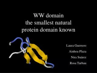
WW domain the smallest natural protein domain known. Laura Guerrero Ainhoa Plaza Nira Suárez Rosa Turbau. Loop 2. Pro (P). Trp (W). Loop 1. WW domain. Mitotic rotamase PIN1. ( Homo sapiens ). WW domain. PIN1. (Human). Structure. only 35-40 aa Monomeric stable in the absence

E N D
WW domainthe smallest natural protein domain known Laura Guerrero Ainhoa Plaza Nira Suárez Rosa Turbau
Loop 2 Pro (P) Trp (W) Loop 1 WW domain. Mitotic rotamase PIN1. (Homo sapiens) WW domain. PIN1. (Human) Structure • only 35-40 aa • Monomeric • stable in the absence of disulfide bonds • three-stranded antiparallel beta-sheet • 2 loops • WWP = 2 tryptophans (20-23aa) + 1 proline.
Dystrophin. (Homo sapiens) P IN1. (Homo sapiens) Function • adaptor module • functional similarity with SH3 domains • attach the enzyme enhancing the catalytic activity • found in many different proteins often localized in the cytoplasm as well as in the cell nucleus. • Proteins participating in signalling paths and development • binds proline-motifs, [X]-P-P-[X]-Y, and/or phosphoserine- phosphothreonine-containing motifs.
Classification Liddle’s sindrome Muscular distrophies Alzheimer disease
WW domain family Sequence Alignment (Clustal W)
COMANDS • ‘ww_totes.fasta’ file: • 1PIN.fa, 1f8aB.fa, 1I8H1.fa, 1i6c1.fa, 1nmv1.fa, 1e0l1.fa, 1eg3A.fa, 1eg4A.fa, 1i5h1.fa, 1e0n1.fa, 1jnq1.fa, 1k9r1.fa, 1k9q1.fa, 1k5r1.fa, 1o6w1.fa, 1tk71.fa • $ clustalw ww_totes.fasta • Outputs are: ww_totes.aln • ww_totes.dnd
Group I. Clustal alignment Group II-III. Clustal alignment
Comandes: ClustalW alignments. WW domain Groups: Group I fileGroupI.fasta {1eg3A.fa, 1eg4A.fa, 1i5h1.fa, 1jmq1.fa, 1k5r1.fa, 1k9q1.fa, 1k9r1.fa} $clustalw Input: fileGroupI.fasta Output: fileGroupI.aln, fileGroupI.dnd Group II-III fileGroupII-III.fasta {1e0l1.fa, 1e0n1.fa, 1o6w1.fa, 1tk71.fa} $clustalw Input: fileGroupII-III.fasta Output: fileGroupII-III.aln, fileGroupII-III.dnd Group IV fileGroupIV.fasta {1f8aB.fa, 1i6c1.fa, 1i8g1.fa, 1I8H1.fa, 1nmv1.fa, 1pinA.fa} $clustalw Input: fileGroupIV.fasta Output: fileGroupIV.aln, fileGroupIV.dnd
WW domain family Sequence Alignment (Clustal W)
WW domain family Sequence Alignment (Clustal W) 1 2 3
Sequence Alignment Native proteins
COMANDS • ‘nativas.fasta’ file: • 1nmv1.fa, 1e0l1.fa, 1e0n1.fa, 1o6w1.fa, 1tk71.fa • $ clustalw nativas.fasta • Outputs are: nativas.aln • nativas.dnd
3 Structure Alignment normal STAMP Cluster: 4 ( 1nmv & 1tk7 1o6w 1e0l 1e0n ) Sc 1.43 RMS 1.62
COMANDS • ‘nativas_stamp.domains’ file: • $ pdbc -d 1nmv1.pdb >nativas_stamp.domains $ pdbc -d 1o6w.pdb >> nativas_stamp.domains $ pdbc -d 1tk7.pdb >> nativas_stamp.domains $ pdbc -d 1e0l1.pdb >> nativas_stamp.domains $ pdbc -d 1e0n1.pdb >> nativas_stamp.domains • $ stamp -l nativas_stamp.domains -rough -n 2 -prefix nativas_stamp > nativas_stamp.out • $ aconvert -in b -out c <nativas_stamp.4> nativas_stamp.aln
3 Structure Alignment advanced STAMP Cluster: 4 ( 1nmv & 1tk7 1o6w 1e0l 1e0n ) Sc 1.11 RMS 1.72
COMANDS • aconvert -in c -out b <nativas.aln> nativas.align • alignfit -f nativas.align -d nativas_stamp.domains -out nativas_stamp_a.trans • stamp -l nativas_stamp_a.trans -prefix nativas_stamp_a > nativas_stamp_a.out
RMSD table 1 2 3 4 5 1 1.08 2.43 0.98 1.75 2 0.00 2.78 1.30 1.51 3 0.00 0.00 2.05 3.27 4 0.00 0.00 0.00 2.02 5 0.00 0.00 0.00 0.00 Structure Alignment XAM
COMANDS: • $ /disc9/Superposition/xam/xam • Input files: 1nmv.pdb, 1o6w.pdb, 1tk7.pdb, 1e0l1.pdb, 1e0n1.pdb Outputs are: nativas_xam.out nativas_xam.pdb
Structure Alignment XAM Loop II 3 TRP (W) 2 TYR (Y) 1 Loop I X-P groove
Structure Alignment XAM Loop I TRP (W) Loop II PRO (P)
Structure Alignment XAM Loop I 3 2 TRP (W) 1 ASN (N) Loop II
1 2 ASN (N) 3 PRO (P) Loop II TRP (W) Structure Alignment XAM Loop I 2
GROUP IV PIN1
PIN1 > Introduction • Phosphorilation-dependent regulation enzime. • Implicated in multiple aspects of cell cycle regulation. • p53, Myt1, Wee1, Cdc25 have been identified as Pin1 interacting proteins. • tau, in neuronal CKS, hyper-phosphorylated in Alzheimer's disease. • Conserved from yeast to human. • Its WW domain is a IV class domain: bind peptides containing a proline residue preceded by a phosphoserine or a phosphothreonine (pSer/pThr –Pro motif)
Structure N-terminal WW domain
Three stranded β-sheet Loop I Loop II Structure > WW domain
Trp29 Tyr18 Structure > WW domain > interacting residues These two residues form an hydrophobic area on the molecular surface of the WW domain
But... Structure > WW domain > interacting residues Pin1’s WWdomain binding site implicates the conservedresiduesTyr18and Trp29which constitute an hydrophobic groove. This hydrophobic binding site alone is not likely to explain the phospho-dependent character of the ligand binding to the Pin1 WW domain.
Hydrogen bonds Structure > WW domain > interacting residues 1st: Phosphate binding pocket: - side chains ofSer11andArg12 - the backbone amide ofArg12 Phosphorilated residue
Arg12 Ser11 Structure > WW domain > interacting residues Located in loop I
Structure > WW domain > interacting residues 2nd: Hibrophobic groove: - Tyr18 - Trp29 constrain the proline at position +1 of the interacting phosphoserine
Trp29 Tyr18 Structure > WW domain > interacting residues
Structure > WW domain > interacting residues Arg12 Ser11 Trp29 Tyr18 Important residues in ligand interaction
Pro32 Pro32 Trp6 Trp6 Structure > WW domain > Structural residues Distance = 2.80 Å
Trp29 Tyr18 Structure > WW domain > Structural residues The hydrophobic groove formed by Trp29 and Tyr18 has a dual role Distance = 3.04 Å
Interaction between Pin1 and its ligands Microtubule-associated tau (τ) protein is a Pin1 WW domain ligand
pThr7 Pro8 Pro9 Interaction between Pin1 and its ligands
pThr7 Ser11 Pro8 Pro9 Arg12 Interaction between Pin1 and its ligands
Ser11 pThr7 pThr7 Arg12 Arg12 Interaction between Pin1 and its ligands Distance = 2.77 Å
Interaction between Pin1 and its ligands Distance = 7.74 Å H bond via phosphate! pThr7 Ser11
Trp29 Trp29 Tyr18 Tyr18 Interaction between Pin1 and its ligands
Structure > WW domain > interacting residues • Arg12 and Ser11 side chains (loop I) anchor the interacting phosphate moiety via several hydrogen bonds. Form the phosphate binding pocket. • The side chain of aromatic Trp29 makes an hydrophobic interaction with the conserved Proline at position +1 of the interacting phosphothreonine and with hydrophobic residue at +2 (Pro9 en tau peptide). • Indeed, Tyr18 makes no interaction with the phosphorilated ligand. • Phosphopeptide ligands are principally fixed by a charge-charge interaction and a proline-aromatic stacking.
Interaction between Pin1 and its ligands Intermolecular stacking structure between Trp29 and Prolines at positions +1 and +2 is analogous to the intramolecular stacking between conserved residues Trp6 and Pro32 in the WWdomain. By mutagenesis studies importance of the Trp6-Pro32 interaction for the folding and the stability of WW domains. Similar strong intermolecular Trp-Pro interaction is used to promote protein-protein interactions between the WW domain and its substrates.
Pro9-Trp29 interaction Pro32-Trp6 interaction Interaction between Pin1 and its ligands