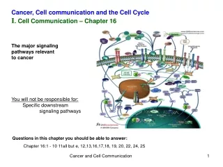
Cell Communication Pathways: Local vs Long-Distance Signaling Mechanisms
Explore the intricate communication systems within cells through local paracrine signaling and long-distance synaptic and hormonal signaling pathways. Discover how growth factors, hormones, and neurotransmitters regulate cellular activities, including cancer development and fruit ripening. Delve into the three stages of cell signaling: reception, transduction, and response, involving key elements like ligands, receptors, G proteins, and second messengers. Unravel the cellular responses triggered by signal-transduction pathways, such as the breakdown of glycogen by epinephrine.
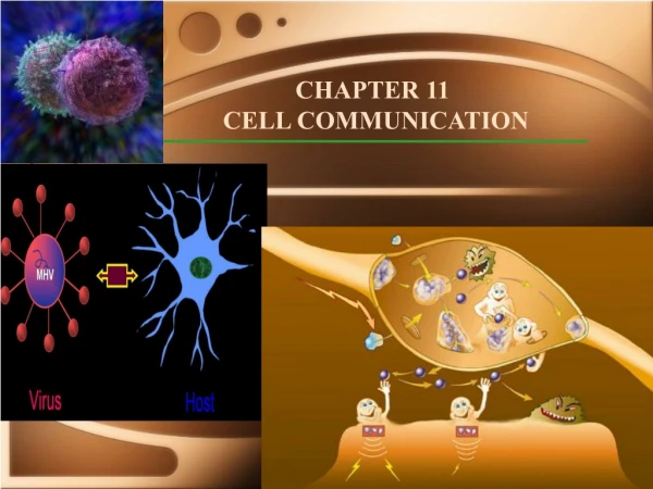

Cell Communication Pathways: Local vs Long-Distance Signaling Mechanisms
E N D
Presentation Transcript
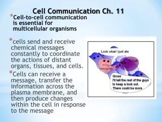
Types of cell talk • Local ‘talk’: Paracrine Signaling (local secretions from neighboring cells), direct cell to cell ‘talk’/via diffusible substances. Ex: growth factors, ethylene gas (plants) • Long-distance ‘talk’: Synaptic (electrical or chemical neuron ‘talk’) signaling, Hormones released into the blood by endocrine glands (ex: epinephrine).
Growth Factors - increase cell division; excess = cancer • Local ‘talk’: Paracrine Signaling (local secretions from neighboring cells), direct cell to cell ‘talk’ via diffusible substances. Ex:growth factors, ethylene gas (plants) • Long-distance ‘talk’: Synaptic (electrical or chemical neuron ‘talk’) signaling, Hormones released into the blood by endocrine glands (ex: epinephrine). Cancer
Ethylene gas = ripening of fruit • Local ‘talk’: Paracrine Signaling (local secretions from neighboring cells), direct cell to cell ‘talk’/via diffusible substances. Ex:growth factors, ethylene gas (plants) • Long-distance ‘talk’: Synaptic (electrical or chemical neuron ‘talk’) signaling, Hormones released into the blood by endocrine glands (ex: epinephrine).
Synaptic talk involves release of neurotransmitters • Local ‘talk’: Paracrine Signaling (local secretions from neighboring cells), direct cell to cell ‘talk’/via diffusible substances. Ex: growth factors, ethylene gas (plants) • Long-distance ‘talk’: Synaptic (electrical or chemical neuron ‘talk’) signaling, Hormones released into the blood by endocrine glands (ex: epinephrine).
Hormones - FIGHT or FLIGHT! • Local ‘talk’: Paracrine Signaling (local secretions from neighboring cells), direct cell to cell ‘talk’/via diffusible substances. Ex: growth factors, ethylene gas (plants) • Long-distance ‘talk’: Synaptic (electrical or chemical neuron ‘talk’) signaling, Hormonesreleased into the blood by endocrine glands (ex: epinephrine).
Epinephrine Hormone Action • One action of Epinephrine = Increase in breakdown of glycogen (polysaccharide) in liver to glucose (why?) • Epinephrine is secreted by adrenal gland (top of kidney), moves through blood, reaches liver • Epinephrine cannot DIRECTLY break down glycogen in a test tube!
The process of epinephrine action (in general cell communication) must involve three stages. 1)Reception - a chemical signal binds to a cellular protein called receptor, typically at the cell’s surface (or in cytoplasm). 2)Transduction- binding leads to a change in the receptor that triggers a series of changes along a signal-transduction pathway 3)Response = a specific cellular activity ex: glycogen breakdown. Fig. 11.5
1) Reception - signal molecule is referred to as “ligand” - usually a small molecule ex: epinephrine/adrenaline Receptor - can be on cell surface (or cytoplasm - ex. steroids). Ligand shape FITS receptor perfectly! (=> SPECIFICITY) Fig. 11.5
2) Receptors can be linked to G proteins • When ligand (ex. Epinephrine) binds to receptor, what follows is SIGNAL TRANSDUCTION (multistep) G proteins are like on-off switches - when GTP is bound = ACTIVE; GDP is bound = INACTIVE
2) G-protein signal transduction system Receptor + Epinephrine = G protein binds to receptor • G protein is activated (GTP attaches). • Activated G protein binds with another membrane protein, (Adenylyl Cyclase - an enzyme). This enzyme is ‘activated’ leading to a cellular response or more signal transduction !
Signal Transduction……. • Adenylyl cyclase converts ATP to cAMP (cyclic AMP). • cAMP is short-lived as another enzyme - phosphodiesterase converts it to AMP (shutting off switch!). • cAMP is referred to as a SECOND MESSENGER Fig. 11.12
Signal Transduction……. More generally, many hormones and other signals trigger the formation of cAMP. • Binding by the signal to a receptor activates a G protein that activates adenylyl cyclase in the plasma membrane. • The cAMP from the adenylyl cyclase diffuses through the cell and activates protein kinaseAwhich phosphorylates other proteins. Fig. 11.13
* Phosphorylation of proteins by a specific enzyme (a protein kinase) is a widespread cellular mechanism for regulating protein activity. Phosphorylation by ‘kinases’ activates (ON), dephoshorylation by ‘phosphatases' inactivates (OFF)! Fig. 11.11
3) Cellular Response - Signal-transduction pathway leads to the regulation of one or more cellular activities. For example, epinephrine helps regulate cellular energy metabolism by activating enzymes that catalyze the breakdown of glycogen. One molecule of epinephrine produces a billion fold increase in the number of phosphorylase enzymes that breakdown glucose. This billion fold increase is possible due to the signal transduction pathway. Also having a multi-step signal ransduction pathway helps control the cellular response better by having different agents fine tune it!
Cellular Response….other ligands act in different ways • Other signaling pathways do not regulate the activity of enzymes but the synthesis of enzymes or other proteins. • These can turn specific genes on or off in the nucleus. Fig. 11.17
Cellular Response…. • Example: Steroids (testosterone) bind directly to 1) receptors in the cytoplasm and this 2) activates proteins (transcription factors) that 3) turn ON/OFF genes! Fig. 11.17
Cellular Response……variations • Epinephrine triggers liver or striated muscle cells to break down glycogen, but cardiac muscle cells are stimulated to contract, leading to a rapid heartbeat.- WHY? And HOW?
Muscle Contraction involves Increase in Calcium Ions - Two of the most important second messengers are cyclic AMP and Ca2+. • Ca2+ concentration is much lower inside than outside the cell. • Various protein pumps transport Ca2+ outside the cell or inside the endoplasmic reticulum or other organelles.
- Signal-transduction pathways trigger the release of Ca2+ from the cell’s ER. • The pathways leading to release involve still other second messengers, diacylglycerol (DAG) and inositol trisphosphate (IP3). • Both molecules are produced by cleavage of certain phospholipids in the plasma membrane.
DAG and IP3 are created when a phospholipase cleaves a membrane phospholipid PIP2. • Phospholipase may be activated by a G protein or a tyrosine-kinase receptor. • IP3 activates a gated-calcium channel, releasing Ca2+. Fig. 11.15
Shutting off G proteins: • The G protein can also act as a GTPase enzyme and hydrolyzes the GTP, which activated it, to GDP. • This change turns the G protein off. • The whole system can be shut down quickly when the extracellular signal molecule is no longer present. Fig. 11.7c
Other types of receptors are not linked to G proteins. Receptors may be tyrosine kinases: • A individual tyrosine-kinase receptor consists of several parts: • an extracellular signal-binding sites, • a single alpha helix spanning the membrane, and • an intracellular tail with several tyrosines. Fig. 11.8a
When ligands bind to two receptors polypeptides, the polypeptides aggregate, forming a dimer. • This activates the tyrosine-kinase section of both. • These add phosphates to the tyrosine tails of the other polypeptide. Fig. 11.8b
The fully-activated receptor proteins activate a variety of specific relay proteins that bind to specific phosphorylated tyrosine molecules. • One tyrosine-kinase receptor dimer may activate ten or more different intracellular proteins simultaneously. • These activated relay proteins trigger many different transduction pathways and responses. Fig. 11.8b
More receptors: • Ligand-gated ion channels are protein pores that open or close in response to a chemical signal. • This allows or blocks ion flow, such as Na+ or Ca2+. • Binding by a ligand to the extracellular side changes the protein’s shape and opens the channel. • Ion flow changes the concentration inside the cell. • When the ligand dissociates, the channel closes.
Cell signaling evolved early in the history of life • Sex is a really important topic of cell talk!
Rather than relying on diffusion of large relay molecules like proteins, many signal pathways are linked together physically by scaffolding proteins. • Scaffolding proteins may themselves be relay proteins to which several other relay proteins attach. • This hardwiring enhances the speed and accuracy of signal transfer between cells. Fig. 11.19