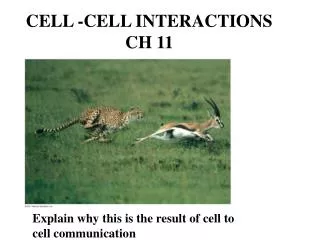
Ch 11 Reading Guide - Cell Communication
820 likes | 845 Vues
Ch 11 Reading Guide - Cell Communication. 1) What is a signal transduction pathway?. ● the process by which a signal on a cell’s surface is converted into a specific cellular response (a series of steps). 2) How do yeast cells communicate while mating?. ● chemical signaling

Ch 11 Reading Guide - Cell Communication
E N D
Presentation Transcript
1) What is a signal transduction pathway? ● the process by which a signal on a cell’s surface is converted into a specific cellular response (a series of steps)
2) How do yeast cells communicate while mating? ● chemical signaling ● 2 mating types: a and α (alpha) -type “a” cells secrete “a” factor -type “α” cells secrete “α” factor ● the factors bind to receptors on the other; the 2 mating factors cause the cells to grow toward each other and fuse
3) How do intercellular connections function in cell to cell communication? ● both plant and animal cells have cell junctions (gap junctions in animal cells; plasmodesmata in plant cells) that, where present, directly connect the cytoplasms of adjacent cells ● signaling substances dissolved in the cytoplasm can freely pass between adjacent cells
4) Explain the two types of local signaling: A) Paracrine signaling ● a secreting cell acts on nearby target cells by releasing molecules of a local regulator (i.e. growth factor) into the extracellular fluid
4) Explain the two types of local signaling: B) Synaptic signaling ● a nerve cell releases neurotransmitter molecules into a synapse, the narrow space between the transmitting cell & the target cell
5) How are long distance signals sent? ● Long distance signals are sent by chemicals called HORMONES. ● specialized endocrine cells secrete hormones into body fluids,often the blood. ● hormones may reach virtually all body cells, but will only attach to target cells with the specific receptor molecule
6) Explain Sutherland’s investigations with epinephrine and the inferences that were derived from this work. ● discovered that epinephrine stimulates glycogen breakdown by activating a cytosolic enzyme, glycogen phosphorylase. ● this only worked when epinephrine was applied to intact cells ● INFERENCE: epinephrine does not act on the enzyme directly; and the cell membrane is somehow involved in transmitting the signal
7) Define the three stages of cell communication: A) Reception: ● the target cell’s detection of a signal coming from outside the cell ● a chemical signal is detected when it binds to a cellular protein (usually a membrane protein)
7) Define the three stages of cell communication: B) Transduction: ● the binding of the signal molecule changes the receptor protein in some way… ● the signal is converted to a form that can bring about a specific cellular response
7) Define the three stages of cell communication: C) Response ● the transduced signal finally triggers a specific cellular response
8) What is a ligand? ● a small molecule that specifically binds to a larger one ● a signal molecule behaves as a ligand
9) What is special about intracellular receptors – hint think of the structure of the cell membrane and how this relates? ● intracellular receptors are typically proteins dissolved in the cytosol or nucleus of a target cell ● may become activated with the binding of the signal molecule ● the activated form may then respond or cause a change (i.e. enter the nucleus and turn on specific genes)
10) Label this diagram of a steroid interacting with an intracellular receptor.(Fig. 11.9)
11) Where would you expect most water soluble messengers to bind and why? ● would most likely bind to receptors on the outside surface of the plasma membrane; ● they are water-soluble & probably too large to pass through the cell membrane
12) What is a G-protein-linked receptor? (see fig. 11.7) ● a plasma membrane receptor that works with the help of a G-protein ● the G-protein is attached to the cytoplasmic side of the membrane and acts as a switch that is on (GTP) or off (GDP)
13) (see fig. 11.7, p. 211 captions): (overview): A G-protein-coupled receptor is a cell-surface transmembrane receptor that works with the help of a G protein, a protein that binds the energy-rich moleculeGTP. (1) When GDP is bound to the G protein, the G protein is inactive. The receptor and G protein work together with another protein, usually an enzyme .
13) (see fig. 11.7, p. 211 captions): (2) When the appropriate signaling molecule binds to the extracellular side of the receptor, the receptor is activated and changes shape . Its cytoplasmic side then binds an inactive G protein, causing a GTP to displace GDP . This activates the G protein.
13) (see fig. 11.7, p. 211 captions): (3) The activated G protein leaves (dissociates from) the receptor, diffuses along the membrane, and then binds to an enzyme, altering the enzyme’sshape & activity. Once activated, the enzyme can trigger the next step, leading to a cellular response.
13) (see fig. 11.7, p. 211 captions): (4) The changes in the enzyme and G protein are only temporary because the G protein also functions as a GTPase enzyme – in other words, it then hydrolyzes its bound GTP to GDP. Now inactive again, the G protein leaves the enzyme, which returns to its original state. The GTPase function of the G protein allows the pathway to shut down rapidly when the signaling molecule is no longer present.
14) What is a KINASE (i.e. a protein kinase)? ● an enzyme that catalyzes the transfer of a phosphate group from ATP to another molecule
15) (see fig. 11.7, p. 212 captions): (overview): Receptor tyrosine kinases belong to a major class of plasma membrane receptors characterized by having enzymatic activity. The part of the receptor protein extending into the cytoplasm functions as a tyrosine kinase, an enzyme that catalyzes the transfer of a phosphate group from ATP to the amino acid tyrosine on a substrate protein. One receptor tyrosine kinase complex may activate ten or more different transduction pathways and cellular responses. The ability of a single ligand-binding event to trigger so many pathways is a key difference between receptor-tyrosine kinases and G protein-coupled receptors.
15) (see fig. 11.7, p. 212 captions): (1) Before the signaling molecule binds, the receptors exist asindividual units referred to as monomers. Each monomer has an extracellularligand-bindingsite, an α helix spanning the membrane, and an intracellular tail containingmultiple tyrosines.
15) (see fig. 11.7, p. 212 captions): (2) Thebinding of a signaling molecule(such as growth factor) causes 2 receptor monomers to associate closely with each other, forming a complex known as a dimer (dimerization).
15) (see fig. 11.7, p. 212 captions): (3) Dimerization activates the tyrosine kinase region of each monomer; each tyrosine kinaseadds a phosphatefrom an ATP molecule to a tyrosine on the tail of the other monomer.
15) (see fig. 11.7, p. 212 captions): (4) Now that the receptor is fully activated, it is recognized by specific relay proteins inside the cell. Each such protein binds to a specific phosphorylated tyrosine, undergoing a resulting structural change that activates the bound protein. Each activated protein triggers atransduction pathway , leading to a cellular response.
16) (see fig. 11.7, p. 213 captions): (overview): What triggers a ligand-gated ion channel to open/close? when a signaling molecule binds as a ligand to the receptor protein What then passes through the channel once it is open?Specific ions, such as Na+ or Ca2+, pass through the channel receptor
16) (see fig. 11.7, p. 213 captions): **study and read the captions for parts 1-3 of this diagram! (conclusion): How do nerve cells make use of ligand-gated ion channels? neurotransmitter molecules released at a synapse between 2 nerve cells bind as ligands to ion channels on the receiving cell, causing the channels to open; as ions flow in/out, an electrical signal is generated and passed down the receiving cell…a nerve impulse!
16) (see fig. 11.7, p. 213 captions): How is a voltage-gated ion channel different?these channels are controlled (opened / closed) by electrical signals (not ligands / chemical signals) .