Chapter 11 Cell communication
Chapter 11 Cell communication. How cells detect, process, and respond to chemical signals send from other cells or from changes in the physical environment ?. Overview: The Cellular Internet Cell-to-cell communication Is absolutely essential for multicellular organisms

Chapter 11 Cell communication
E N D
Presentation Transcript
Chapter 11Cell communication How cells detect, process, and respond to chemical signals send from other cells or from changes in the physical environment ?
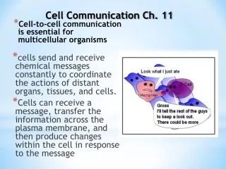
Overview: The Cellular Internet • Cell-to-cell communication • Is absolutely essential for multicellular organisms • External signals are converted into responses within the cell Ex. Yeast cells --- Identify their mates by cell signaling
Exchange of mating factors. Each cell type secretes a mating factor that binds to receptors on the other cell type. factor 3 1 2 Receptor a factor Yeast cell, mating type a Yeast cell, mating type Mating. Binding of the factors to receptors induces changes in the cells that lead to their fusion. a New a/ cell. The nucleus of the fused cell includes all the genes from the a and a cells. a/ Cell signaling evolved early in the history of life Saccharomyces cerevisiae“yeast” --- identify their mates by chemical signaling Two sex,(mating type) Without actually entering the cells, the receptor-bound molecules of the two mating factors cause the cells to grow toward each other and bring about other cellullar changes. Figure 11.2
The process by which a signal on a cell’s surface is converted into a specific cellular response is a series of steps --- called a signal-transduction pathway • Signal transduction pathways • Convert signals on a cell’s surface into cellular responses • Are similar in microbes and mammals, suggesting an early origin • Cells in a multicellular organism --- communicate via chemical messengers
Plasma membranes Plasmodesmata between plant cells Gap junctions between animal cells Figure 11.3 (a) Cell junctions. Both animals and plants have cell junctions that allow molecules to pass readily between adjacent cells without crossing plasma membranes. • Animal and plant cells • Have cell junctions that directly connect the cytoplasm of adjacent cells
Figure 11.3 (b) Cell-cell recognition. Two cells in an animal may communicate by interaction between molecules protruding from their surfaces. • In local signaling, animal cells • May communicate via direct contact
Local signaling Electrical signal along nerve cell triggers release of neurotransmitter Target cell Neurotransmitter diffuses acrosssynapse Secretory vesicle Local regulator diffuses through extracellular fluid Target cell is stimulated (a) Paracrine signaling. A secreting cell acts on nearby target cells by discharging molecules of a local regulator (a growth factor, for example) into the extracellular fluid. (b) Synaptic signaling. A nerve cell releases neurotransmitter molecules into a synapse, stimulating the target cell. Figure 11.4 A B Paracrine signaling: --- the transmitting cell secretes molecules of a local regulator a substance that influences cells in the vicinity. • In other cases, animal cells • Communicate using local regulators Growth factor neurotransmitter
* Endocrine signaling: (animal cells) --- specialized cells release hormone molecules into vessels of the circulatory system. * In plants, hormones sometimes travel in vessels but more often reach their targets by moving through cells or by diffusion through the air as a gas. • In long-distance signaling • Both plants and animals use hormones Long-distance signaling Blood vessel Endocrine cell Hormone travels in bloodstream to target cells Target cell The plant hormone ethylene, a gas that promotes fruit ripening and helps regulate growth. (C2H4) (c) Hormonal signaling. Specialized endocrine cells secrete hormones into body fluids, often the blood. Hormones may reach virtually all body cells. Figure 11.4 C
What happens when a cell encounters a signal ? The signal must be recognized by a specific receptor molecule, and the information it carries must be changed into another form-transduced-inside the cell before the cell can respond.
The three stages of cell signaling are reception, transduction, response Earl W. Sutherland (Nobel Prize in 1971) How the animal hormone epinephrine stimulates breakdown of the storage polysaccharide glycogen within liver and skeletal muscle cells. Glycolysis: Glycogen Glucose-1-phosphate Glucose-6-phosphate glucose
Glycogen Glucose-1-phosphate Glucose-6-phosphate glucose Epinephrine Enzyme: Glycogen phosphorylase • When epinephrine was added to a • Test-tube mixture containing the • phosphorylase and its substrate, glycogen • no depolymerization occurred • 2. Epinephrine could activate glycogen phosphorylase • only when it was added to a solution containing • intact cells.
Epinephrine does not interact directly with the enzyme responsible for glycogen breakdown. • 2. The plasma membrane is somehow involved in transmitting the epinephrine signal. • Catalysis by an enzyme • Rearrangement of the cytoskeleton • Activation of specific genes in the nucleus Relay molecules
Signal reception and the initiation of transduction a yeast cell only “heard” the signals by its prospective mates, a cells. The signal receptor is the identity tag on the target cell. A signal molecule binds to a receptor protein, causing the protein to change shape. ligand * The term for a small molecule that specifically binds to a larger one Key & lock receptor Conformational change activation receptor
Most signal receptors are plasma membrane proteins Receptor transmits information from the extracellular environment to the inside of the cell by changing shape or aggregating when a specific ligand binds to it. Three major types of membrane receptors: 1.G-protein-linked receptors 2. tyrosin-kinase receptors 3. ion-channel receptors
G-protein-linked receptors • --- a plasma membrane receptor • --- works with the help of a protein called a G protein • --- vary in their binding sites for recognizing signal • molecules and for recognizing different G proteins
Signal-binding site Segment that interacts with G proteins Inctivate enzyme ActivatedReceptor G-protein-linked Receptor Signal molecule Plasma Membrane GDP G-protein(inactive) GTP GDP CYTOPLASM Enzyme Activated enzyme GTP GTP GDP GDP Pi Pi Cellular response Cellular response G-protein-linked receptor: --- widespread and diverse in functions. * mouse embryogenesis * sensory reception (vision and smell) 視覺 嗅覺 --- involved in diseases. cholera 霍亂 pertussis 百日咳 G protein: GDP bound --- inactive GTP bound --- active 1. 2. Figure 11.7 3. 4. Figure 11.7
Signal-binding sitea Signalmolecule Signal molecule Helix in the Membrane Tyr Tyr Tyr Tyr Tyrosines Tyr Tyr Tyr Tyr Tyr Tyr Tyr Tyr Receptor tyrosinekinase proteins(inactive monomers) Dimer CYTOPLASM Activatedrelay proteins Cellularresponse 1 P P Tyr Tyr Tyr Tyr P Tyr P Tyr Tyr Tyr P P P Tyr Tyr Tyr Tyr Tyr P Tyr Tyr Tyr Cellularresponse 2 P P P Tyr Tyr Tyr Tyr Tyr P 6 ATP 6 ADP Tyr Tyr Tyr Activated tyrosine- kinase regions (unphosphorylated dimer) Fully activated receptor tyrosine-kinase (phosphorylated dimer) Inactiverelay proteins 2. Tyrosine-Kinase Receptors --- a type of receptor specialized for triggering more than one signal-transduction pathway at once. ( have enzymatic activity 酵素活性: tyrosin kinase) • Polypeptide aggregation • Phosphorylation of the receptor
The ability of a single ligand-binding event to trigger so many pathways is a key difference between these receptors and G-protein-linked receptors. Ligand independent activation of tyrosin-kinase receptor Mutation (突變) Cancer
Gate closed Signalmolecule(ligand) Ions Ligand-gated ion channel receptor Plasma Membrane Gate open Cellularresponse Gate close Figure 11.7 Ion-channel receptors: Ligand-gated ion channels * Nervous system
Not all signal receptors are membrane proteins !! Intracellular receptors : --- in the cytosol or nucleus of target cells. --- * hydrophobic : steroid hormones tyroid hormones ex. Testosterone (one steroid hormone) --- secreted from testis * small molecules : nitric oxide (NO)
Activated testosterone receptor Transcription factors In nucleus: ex, estrogen receptors transcription translation
Signal-transduction pathways Relay molecules • Catalysis by an enzyme • Rearrangement of the cytoskeleton • Activation of specific genes in the nucleus --- multistep pathway --- signal amplification Protein phosphorylation Conformational change
Protein phosphorylation: --- a widespread cellular mechanism for regulating protein activity. Protein phosphatase: (去磷酸酶) - an enzyme that remove phosphate groups from proteins. Protein kinase: (磷酸激酶) - an enzyme that transfers phosphate groups from ATP to a protein. ATP ADP kinase A A P phosphatase active inactive P i
Protein kinase: tyrosin kinase serine/threonine kinase phosphatase kinase 1% A phosphorylation cascade
Not all components of signal-transduction pathway are proteins !! Many signaling pathways also involve small, nonprotein, water-soluble molecules or ions Second messengers * Spread by “diffusion” * Participate in pathways initiated by both G-protein-linked receptors and tyrosin-kinase receptors. * Including cyclic AMP and calcium ions (Ca2+).
NH2 NH2 NH2 N N N N N N N N N N N O O O N O Adenylyl cyclase Phoshodiesterase CH2 O HO Ch2 P –O O P O P P O CH2 O O O O O O O O O P Pyrophosphate H2O O O P P i OH OH OH OH OH ATP Cyclic AMP AMP Figure 11.9 Cyclic AMP Cyclic adenosine monophosphate; cyclic AMP; cAMP
First messenger (signal molecule such as epinephrine) Adenylyl cyclase G protein GTP G-protein-linked receptor ATP cAMP Protein kinase A Cellular responses • Many G-proteins • Trigger the formation of cAMP, which then acts as a second messenger in cellular pathways * Some are inhibitory G protein which inhibit adenylyl cyclase. * Cholera --- Vibrio cholerae --- produce a toxin, which modifies a G protein involved in regulating salt and water secretion. Figure 11.10
Neurotransmitters Growth factors hormones Ca2+ More widely used than cAMP as a second messenger Cytosolic concentration of calcium ions (Ca2+) Muscle cell contraction secretion Cell division
EXTRACELLULAR FLUID Plasma membrane Ca2+pump ATP Mitochondrion Nucleus CYTOSOL Ca2+pump Endoplasmic reticulum (ER) ATP Ca2+pump Key High [Ca2+] Low [Ca2+] Figure 11.11 Ca2+ concentration in the cytosol is normally much lower than the concentration outside the cell. 10,000X * Diacylglycerol (DAG) * Inositol trisphosphate (IP3)
3 4 6 5 2 1 * Calcium and IP3 in signaling pathways. A signal molecule binds to a receptor, leading to activation of phospholipase C. DAG functions as a second messenger in other pathways. Phospholipase C cleaves a plasma membrane phospholipid called PIP2 into DAG and IP3. EXTRA- CELLULAR FLUID Signal molecule (first messenger) G protein DAG GTP PIP2 G-protein-linked receptor Phospholipase C IP3 (second messenger) IP3-gated calcium channel Endoplasmic reticulum (ER) Various proteins activated Cellularresponse Ca2+ Ca2+ (second messenger) IP3 quickly diffuses through the cytosol and binds to an IP3– gated calcium channel in the ER membrane, causing it to open. The calcium ions activate the next protein in one or more signaling pathways. Calcium ions flow out of the ER (down their con- centration gradient), raising the Ca2+ level in the cytosol. Figure 11.12
Reception Growth factor Reception Binding of epinephrine to G-protein-linked receptor (1 molecule) Receptor Transduction Inactive G protein Active G protein (102 molecules) Phosphorylation cascade Transduction Inactive adenylyl cyclase Active adenylyl cyclase (102) CYTOPLASM ATP Cyclic AMP (104) Inactive protein kinase A Active protein kinase A (104) Inactive transcription factor Active transcription factor Inactive phosphorylase kinase Response Active phosphorylase kinase (105) P DNA Inactive glycogen phosphorylase Active glycogen phosphorylase (106) Gene Response Glycogen mRNA Glucose-1-phosphate(108 molecules) NUCLEUS Concept 11.4 Response: cell signaling leads to regulation of cytoplasmic activities or transcription cytoplasmic response nucleus response
Reception Binding of epinephrine to G-protein-linked receptor (1 molecule) Transduction Inactive G protein Active G protein (102 molecules) Inactive adenylyl cyclase Active adenylyl cyclase (102) ATP Cyclic AMP (104) Inactive protein kinase A Active protein kinase A (104) Inactive phosphorylase kinase Active phosphorylase kinase (105) Inactive glycogen phosphorylase Active glycogen phosphorylase (106) Response Glycogen Glucose-1-phosphate(108 molecules) • Two important benefits: • Signal amplification • The specificity of response Why are there often so many steps between a signaling event at the cell surface and the cell’s response ? * At each catalytic step in the cascade, the number of activated products is much greater than in the preceding step. Figure 11.13
Signaling pathways with a multiplicity of steps have two important benefits: 1. amplify the signal 2. contribute to the specificity of response The specificity of cell signaling epinephrine Liver cell heart cell Glycogen breakdown contraction
Signalmolecule Receptor Relaymolecules Response 1 Response 2 Response 3 Activationor inhibition Response 4 Response 5 The response of a particular cell to a signal depends on its particular collection of signal receptor proteins, relay proteins, and proteins needed to carry out the response. Cell A. Pathway leads to a single response Cell B. Pathway branches, leading to two responses Cell D. Different receptor leads to a different response Cell C. Cross-talk occurs between two pathways Figure 11.15
Signaling Efficiency: Scaffolding Proteins and Signaling Complexes • Scaffolding proteins • Can increase the signal transduction efficiency Signalmolecule Plasmamembrane Receptor Threedifferentproteinkinases Scaffoldingprotein Figure 11.16
A key to a cell’s continuing receptiveness to regulation is the reversibility of the changes that signals produce. • Signal response is terminated quickly • By the reversal of ligand binding • The relay molecules return to their inactive forms. Termination of the Signal How ?