Download
1 / 89
890 likes | 1.41k Vues
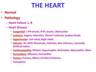
Heart murmurs & Dynamic Auscultation. Dr Nithin P G. Outlay of Seminar. Definition What to look for/ how to describe a murmur Classification of murmurs Types of murmurs Dynamic Auscultation. Definition of murmur. “Relatively prolonged series of audible vibrations”

E N D
Heart murmurs & Dynamic Auscultation Dr Nithin P G
Outlay of Seminar • Definition • What to look for/ how to describe a murmur • Classification of murmurs • Types of murmurs • Dynamic Auscultation
Definition of murmur • “Relatively prolonged series of audible vibrations” • Characterized by the timing in cardiac cycle, intensity (loudness), frequency (pitch), quality, configuration, duration and direction of radiation.
How is a murmur produced? Sound is produced by vibration Turbulence generated in the blood column vibrations set up in the vessel wall & cardiac structures murmurs
L = linear dimension (internal diameter In pipes) V = mean fluid velocity Q = volumetric flow rate A = pipe cross-sectional area m = dynamic viscosity of the fluid n = kinematic viscosity [m/ r] r = density of the fluid Q = V1*A1= V2*A2 Q = P/R How is a murmur produced? [Re >4000 turbulent flow] Re => Turbulence => murmur
Auscultation of murmur Other factors affecting auscultation of murmur • Distance from chest wall, position of patient • Underlying soft tissue, lung, fluid • Quality of apparatus
Auscultation of murmur Upper 3rd L St-C Jn 4th L cos. cart. in L St. border 4th RICS
Description of a Murmur • Position in the cardiac cycle • Site of murmur [max. intensity] • Intensity • Quality & Pitch • Conduction • Dynamic changes
Position in the cardiac cycle early systolic mid systolic • Systolic murmur late systolic pan/holo systolic early diastolic • Diastolic murmur mid diastolic pre systolic • Continuous murmur
Intensity- Grading FREEMAN & LEVINE GRADING GRADE 1- faintest murmur which can be heard only with special effort. GRADE 2- soft but readily audible GRADE 3- loud without thrill GRADE 4- loud with thrill GRADE 5- heard with steth partially off the chest GRADE 6- heard with steth held off the chest wall.
Quality & Pitch • Depends on two factors • Pressure difference or gradient- Gr pitch • Amount of Flow- Flow pitch
Conduction of murmur Site to which conducted aids in diagnosis • MS localized to apex • MR conducted to axilla and back; LLSB in MVP-MR • AS conducted to Carotids
Classification & types of murmurs early systolic mid systolic • Systolic murmur late systolic pan/holo systolic early diastolic • Diastolic murmur mid diastolic pre systolic • Continuous murmur
Midsystolic murmur • Most common murmur heard in everyday practice. • Starts at an interval after S1 and ends before S2. • It could be PATHOLOGICAL INNOCENT/PHYSIOLOGICAL • 5 settings • Ventricular outflow obstruction • Dilation of aorta and pulmonary trunk • Accelerated systolic flow into aorta or pulmonary trunk • Innocent midsystolic murmur( including those due to morphological changes of valve with no obstruction) • Some forms of MR
Ventricular outflow obstruction Phasic flow across left and right outflow tract • Isovolumic contraction (b) • Maximal ejection (c) • Start of relaxation and reduced ejection (d) • Isovolumic relaxation (e) • LV filling, rapid phase (f) • Slow LV filling (diastasis) (g) • Atrial systole or booster (a)
IVC S1 ventricular pressure increases opening of Aorta and pulmonary valve ejection commences and murmur begins Ejection increases murmur becomes crescendo Ejection declines murmur in decrescendo Murmur ends before ventricular pressure drops below aortic pressure at which aortic valve and pulmonary valve closes generating a2 and p2 AS
AS • Harsh, crescendo-decrescendo MSM • Early sys peak short duration vs. Late systolic peak long duration • Always Symmetrical [vs. PS] • ES absent in calcific valves, sub and supra valvular AS • Length and loudness do not necessarily corresponds to severity but length more suggestive of severity than other murmurs Reverse splitting S2 S4
AS • Gallaverdin phenomenon/ hourglass phenomenon Lower n (aortic)vs. Higher n (mitral) periodic vibrations of stiffened non calcific aortic valve • Differentiating from MR
AS Postextrasystolic enhancement results from the variable interaction of three factors: • Increase in the contractile state (inotropism) of the ventricular muscle which is more evident if there is hypertrophy and/or depressed ventricular function. • The pause provides longer filling time for the ventricle, which is more consequential in hypertrophic ventricles (e.g., aortic stenosis) than in ventricular volume overload states (e.g., mitral regurgitation). • Lastly, there is more time for arterial runoff, and in the case of aortic regurgitation, more backflow into the ventricle. This effect lowers the arterial diastolic pressure and the impedance to forward flow (afterload) in the beat following the pause.
Dynamic LVOT obstruction Factors increasing gradient LV Contractility Exercise Cathecolamines Digitalis Ventricular Volume Valsalva Standing Nitroglycerine/ Amyl nitrate Tachycardia Aortic impedance and pressure Sustained Handgrip Passive Leg Raise HOCM
PS • Murmur brought on by a phasic ejection click; radiates up & left • As severity increases length increases and P2 becomes soft (abruptness of closure reduced), S2 split widens, S4 • Loses symmetry becomes kite shaped • May engulf A2 and P2 may be inaudible; may be confused with VSD
Other causes of MSM Dilation of Aorta & Pulmonary trunk • Short soft midsystolic murmur • Left sided murmurs in marfan’s syndrome, syphilis • Right sided murmurs in idiopathic dilation of pulmonary artery, pulmonary hypertension MSM of Hyperdynamic circulation • Normal aorta or pulmonary trunk but increased flow • Anaemia, pregnancy, fever, thyrotoxicosis
Other causes of MSM OS-ASD • Rapid flow across pulmonary valve to dilated pulmonary trunk Pure AR • Due to Accelerated LV ejection
Physiological causes Innocent systolic murmur • Still’s murmur • Pulmonary mid systolic murmur • Peripheral pulmonary systolic murmur • Supraclavicular or brachiocephalic systolic murmur • Aortic sclerosis • Systolic mammary soufflé
Still’s murmur Short buzzing murmur ‘twanging of a rubber band’ Pure medium frequency by periodic vibrations of pulmonic leaflets at their attachment Physiological murmurs
Pulmonary mid systolic murmur & Peripheral pulmonary systolic murmur Angulation and disparity between pulmonary trunk and its branches turbulent flow Normally disappears with maturity of pulmonary bed Physiological murmurs
Supraclavicular or brachiocephalic systolic murmur Aortic origins of major normal brachiocephalic arteries Crescendo-decrescendo, abrupt onset, short, sometimes radiating below clavicle vs. supra valvular AS – these murmur are softer below clavicle and decreases with shoulder abduction Physiological murmurs
Mammary Soufflé Late Pregnancy or puerperium Sometimes continuous louder in systole, distinct gap from S1 [ time for ejected blood to reach mammary arteries] 2 or 3 RICS/ LICS Light Pressure augments murmur becomes continuous; firm Pr abolishes murmur Physiological murmurs
Pan Systolic/ Holo Systolic Murmur Flow from a chamber or vessel whose pressure or resistance throughout systole is higher than pressure or resistance of the chamber receiving the flow • Mitral Regurgitation • Tricuspid Regurgitation • Ventricular Septal Defect • Aorto Pulmonary Window • Patent Ductus Arteriosus with PAH
S1 to S2 provided MV remains incompetent and gradient remains Holosystolic Early systolic Late systolic Sometimes MSM Radiates to axilla and back becos jet directed posterolaterally in LA LLSB when jet directed against atrial septum near base of aorta Mitral Regurgitation
LLSB- RA Rivero Carvallo’s sign- Increased VR, increased RV volume Increased SV velocity of regurgitant flow Sometimes TR heard only during inspiration Carvallo’s sign disappears in RV failure Diff from organic TR PSM vs. ESM High n vs. Medium n Features of PAH present Tricuspid Regurgitation
Depends on site, size and gradient Very restrictive VSD- ESM decrescendo pattern Mod and NR VSD- PSM Sub arterial VSD- 1 or 2 LICS similar to PS murmur Septal aneurysms- click with LSM or PSM with late Sys Accentuation Large shunt – MDM NR VSD with PAH- ESM PSM absent in Eissenmenger Syndrome Ventricular Septal Defect
Other PSM • Aorto Pulmonary Window with PAH • Otherwise continuous murmur • Diastolic component reduced with increasing PAH • PDA with PAH • Similar mechanism
Acute Mitral Regurgitation Decrescendo murmur Non distensible LA , large v wave approaching LV pressure in late systole Maximum flow early systole and minimum to nil flow in late systole ESM
Other ESM • Normal pressure TR, Organic TR Tall RA v waves reach the level of normal RV pressure in late systole, so lower rate of regurgitant flow Moderate to low frequency as compared to high frequency in high pressure TR • VSD with PVR or small muscular VSD -Equalization of pressures in cases of PAH -Small VSD closes in late systole
MVP Leaflets remains competent during early ventricular contraction but overshoot in late systole [critical V. dimensions] One or more mid systolic clicks precede murmur [sudden deceleration of the column of blood against the prolapsed leaflet or scallops] Longer and softer Prompt standing after squatting Valsalva II Short & louder squatting Sustained hand grip Amyl nitrate Other LSM- papillary muscle dysfunction Post Pap Muscle . Late systolic cresendo to S2 LSM Barlow’s syndrome refers to the spectrum of symptoms caused by MVP [click or murmur alone to palpitations, chest pain, or syncope]
Early diastolic murmur • AR murmur -Soft high frequency early diastolic murmur with pt sitting & leaning forward in full held expiration -3 LICS [ 2 & 3 RICS in root dil] -musical quality in eversion -Austin Flint murmur -Cole- Cecil murmur- AR murmur in left axilla due to higher position of apex
AR • Difference between acute and chronic AR • Austin Flint Murmur to be discussed
High pitched soft blowing decrescendo murmur usually lasts throughout diastole heard in the left upper sternal border Associated with loud P2 and other features of PAH PR vs. AR Loud P2, murmur begins after P2 Normal pulse pressure Clinical setting Squatting and sustained hand grip increases AR High Pressure PR