Download
1 / 24
240 likes | 1.04k Vues
Hemorrhagic fever DIC Tourniquet test. Hemorrhagic fever. Definition: diverse group of illnesses results from infection with one of several single-stranded RNA viruses (members of the families Arenaviridae , Bunyaviridae , Filoviridae , and Flaviviridae ). . Flaviviruses cause?

E N D
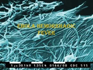
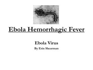
Hemorrhagic fever DIC Tourniquet test
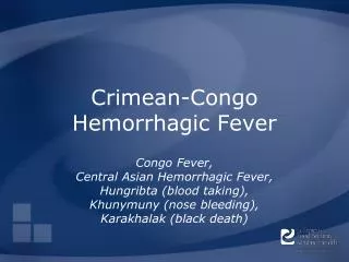
Hemorrhagic fever • Definition: diverse group of illnesses results from infection with one of several single-stranded RNA viruses (members of the families Arenaviridae, Bunyaviridae, Filoviridae, and Flaviviridae).
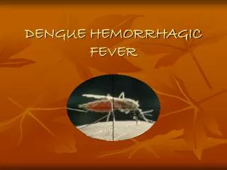
Flaviviruses cause? • Dengue and yellow fever
What are these viruses? • They are all RNA viruses, and all are covered, or enveloped, in a fatty (lipid) coating. • Their survival is dependent on an animal or insect host, called the natural reservoir. • The viruses are geographically restricted to the areas where their host species live. • Humans are not the natural reservoir for any of these viruses. Humans are infected when they come into contact with infected hosts • With a few noteworthy exceptions, there is no cure or established drug treatment for VHFs.
Who are the hosts? • Zoonotic
General clinical features • Incubation period: 2 days for the Rift Valley fever or as long as 21 days for Lassa fever • Early phase: early phase of a viral hemorrhagic fever are very similar, irrespective of the causative virus, and resemble a flu-like illness or gastroenteritis. Hepatitis is common. • The late phase is more specific and is characterized by organ failure, persistent leukopenia, altered mental status, and hemorrhage. Exanthemas (rash) and mucosal lesions can occur.
On examination • On examination patients may have conjunctival injection, mild hypotension, flushing, and petechial hemorrhages. Bleeding is variable and generally not life threatening, but it is an index of severity
How do the viruses cause these symtoms? • VHF viruses target vascular endothelium, causing microvascular damage and derangement in vascular permeability.
Laboratory findings • Thrombocytopenia, leukopenia, anemia, increased hematocrit, elevated liver function tests, and findings consistent with disseminated intravascular coagulation. • Urinalysis can reveal proteinuria and hematuria.
Prevention is the key! • With the exception of yellow fever and Argentine hemorrhagic fever, for which vaccines have been developed, no vaccines exist that can protect against these diseases. • Therefore, prevention efforts must concentrate on avoiding contact with host species.
Treatment • Patients receive supportive therapy, but generally speaking, there is no other treatment or established cure for VHFs. • Ribavirin, an anti-viral drug, has been effective in treating some individuals with Lassa fever or HFRS (Hemorrhagic fever with renal syndrome is a group of clinically similar illnesses caused by hantaviruses from the family Bunyaviridae.)
From Principles of Critical Care: Chapter 60 - Viral Hemorrhagic Fevers via Access Medicine
What is DIC? • Disseminated intravascular coagulation (DIC) is an acquired coagulation disorder resulting from excessive activation of the coagulation system. • Arises from? • sepsis, massive tissue injury, and obstetric complications being amongst the most common. • DIC is always secondary, and increases the risk of mortality beyond that associated with the primary disease.
What are the 5 components? • Exposure of blood to procoagulants • Formation of fibrin in the circulation • Fibrinolysis • Depletion of clotting factors • End-organ damage
Tissue factor • In response to cell injury or activation, monocytes and endothelial cells generate tissue factor activates the coagulation cascade. • In DIC, the normal anticoagulant and fibrinolytic systems are overwhelmed and coagulation activation cannot be contained. • The process rapidly becomes systemic, resulting in disseminated microvascularthrombi.
What is increased in this state? • Bleeding • Coagulation factors and natural anticoagulants are consumed during thrombosis, as are platelets, all of which become depleted.
The fibrinolytic system is activated to dissolve the fibrin thrombi. • Therefore, plasminogenis consumed as it is converted into plasmin, which in turn breaks down fibrin clots. • Fibrin degradation products (FDP) including D-dimers are formed, which can contribute to bleeding, because they impair fibrin clot formation and interfere with platelet function
The pathogenesis of acute DIC is mediated by the widespread release of thrombin and plasmin into the circulation. Tissue factor is released from damaged tissue, leading to unregulated thrombin formation.
What is chronic DIC? • Compensated or chronic DIC develops when blood is continuously or intermittently exposed to small amounts of tissue factor and compensatory mechanisms in the liver and bone marrow are largely able to replenish the depleted coagulation proteins and platelets, respectively. • Under these conditions, the patient is either asymptomatic with increased levels of fibrin degradation products or has manifestations of venous and/or arterial thrombosis. • Patients with chronic DIC may also have minor skin and mucosal bleeding
Treatment • Treatment is primarily that of the underlying cause, together with supportive measures as necessary (e.g. plasma or platelet replacement)
Tourniquet test • The tourniquet test reflects both capillary fragility and thrombocytopenia. • Increased vascular permeability, bleeding, and possible DIC may be mediated by circulating dengue antigen-antibody complexes, activation of complement, and release of vasoactive amines. • For dengue fever - • Study 1 – Malaysia • The sensitivity and specificity of the tourniquet test was 82.8% and 23.5% respectively. • Study 2 – India • The tourniquet test was positive in only 39.1% of all DHF cases.
It seems that in a hospital setting, the tourniquet test adds little to the diagnosis of dengue infection/DHF. • But may be useful in diagnosing dengue infection in busy rural health stations in dengue endemic areas of the tropics.