Download
1 / 31
310 likes | 330 Vues
Learn about the structure of the cell membrane and how molecules are selectively transported across it. Understand the role of phospholipids, proteins, and cholesterol in membrane function. Explore the fluid mosaic model and the importance of surface area to cell volume ratio.

E N D
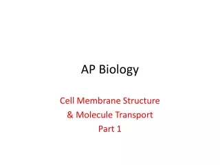
AP Biology Cell Membrane Structure & Molecule Transport Part 1
Selectively Permeable-The cell “selects” what materials enter or exit the cell through the membrane
Membrane Structure • Phospholipids make up the majority of the cell membrane and also organelle membranes. • These are Amphipathic Molecule. (It means there is a hydrophilic and hydrophobic component.) • These molecules create the bi-layer and the structure is held intact by the presence of water outside and inside the cell. The negatively charged phosphorus line up to make a barrier preventing water from forming hydration shells, water surrounding a molecule, around the phospholipids and thereby dissolving the membrane.
AmphipathicPhospholipids WATER Hydrophilic head Hydrophobic tail WATER
Hydrophilic region of protein AmphipathicProteins Phospholipid bilayer Hydrophobic region of protein
Proteins • These are also Amphipathic molecules. (This is due to proteins folding into a 3-D structure and that proteins are composed of amino acids, of which some are polar and some are non-polar.)
Two types of proteins are present on membranes: • Integral – These run completely through the bi-layer from the outside to the inside. • These function in the transport of molecules and foundation. (Help to maintain the INTEGRITY of the structure.) • Peripheral – These are located on one side of the membrane. (They do not extend into the bi-layer of the membrane. • These act as sites for attachment of the Cytoskeleton on the inside of the cell and the attachment of the Extra Cellular Matrix, ECM, (like armor for the fragile cell) on the outside of the cell.
The proteins of the cell membrane can have several functions. • Molecule transport (Helps move food, water, or something across the membrane.) • Act as enzymes (To control metabolic processes.) • Cell to cell communication and recognition (So that cells can work together in tissues.) • Signal Receptors (To catch hormones or other molecules circulating in the blood.) • Intercellular junctions (For “stiching” cells together to make tissues.) • Attachment points for the cytoskeleton and ECM
Signal Enzymes Membrane Protein Functions Receptor ATP Enzymatic activity Transport Signal transduction
Membrane Protein Functions Glyco- protein Attachment to the cytoskeleton and extra- cellular matrix (ECM) Cell-cell recognition Intercellular joining
Cholesterol • This molecule helps keeps the cell membrane flexible to some degree. • It also helps to keep the cell membrane of plant cells from freezing solid in very cold temperatures, like the Tundra.
The membrane is described as a Fluid-Mosaic model because it looks like a moving (Fluid) puzzle (mosaic). • All the pieces can move laterally, like students moving from seat to seat. The proteins moving in this sea of phospholipids would be like the teacher moving around the student desks. Imagine the ceiling and floor are water molecules. They keep you from moving up and down to some extent by their presence. • Scientific Model • These are used to represent what is difficult to actually see. (Like a model of the solar system. or the model of DNA or a cell membrane.
Fluid Mosaic Model and Phospholipids Lateral movement (~107 times per second) Flip-flop (~ once per month) Movement of phospholipids
The Importance of Surface Area to Cell Volume relationship • All cells are considered Open Systems in their natural settings because there are materials coming into the cell from the surrounding environment; as well as, materials leaving the cell and going into the surrounding environment. The cell is open to interaction with the environment. • Remember, cells can only be so small. (There has to be ENOUGH room (volume) to hold things and to perform work inside a cell using the cell membrane.) • Cells can only be so large. ( Larger means more traffic going in both directions across the cell membrane)
A cell must be large enough to contain DNA and Ribosomes for making proteins, and some cytoplasm to act as working “space”. They can only be so big because we have to be able to move enough “Food” into and “waste” out of a cell efficiently. If it is too large the cell becomes inefficient at moving these things so it divides to get back to a smaller state
How do you increase surface area WITHOUT increasing cell volume significantly? • ANSWER: Folding of the membrane. Look at the following examples: • Your lungs and other respiratory surfaces. • The surface of the cells needs to stay wet for gas exchanges to occur; but notice the increased surface area by folding. This allows for the transport of the massive amounts of oxygen that organisms need to live AS WELL AS the CO2 out as a waste product. • Your lungs have the surface area equal to a tennis court, all jammed into your chest.
If all of the capillaries that surround the alveoli were unwound and laid end to end, they would extend for about 992 kilometers (616 mi).
Your and other animal intestines. Look at the folding at the organ and at the cellular levels. This allows your body to taken in the massive amounts of food we eat. After all, you have a trillion cells to feed and they need food all day and night long. • This section of intestine has finger-like projections, called villi, which increases surface area. • Smaller finger-like membrane projections, called microvilli, are located on the cells covering the villi; this too increases surface area again. • THE TOTAL SURFACE AREA IS EQUAL TO THE SIZE OF A FOOTBALL FIELD!!!!!
Key Nutrient absorption Vein carrying blood to hepatic portal vessel Microvilli (brush border) Villi and Microvilli on the interior of the small intestine Blood capillaries Epithelial cells Muscle layers Epithelial cells Large circular folds Lacteal Villi Lymph vessel Villi Intestinal wall
Your and other animals’ excretory systems. Thousands of cells all try to help purify the liquid component of blood by performing selective molecule transport. The buildup of Nitrogen containing Ammonia (NH3) comes from our cells utilizing proteins for energy production. Our excretory system gets rid of the Ammonia either as straight Ammonia (In the case of fish), as urea (as in most land animals, including humans), or as Uric acid (for birds and reptiles) ALSO, notice that Urea and Uric Acid get rid of an additional waste product… CO2. (Remember, this is part of the Nitrogen Cycle.)