Download

1 / 24
240 likes | 530 Vues
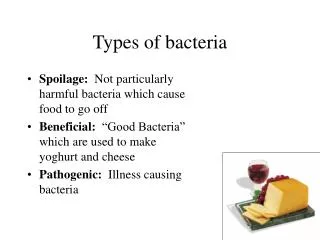
Lab Diagnosis of Bacteria. causing STDs. Prof. Omayma Mohammad. Neisseria gonorrhea Specimen: - In females: cervical swab in acute and chronic infection. In males: urethral discharge in acute infection and morning drops in chronic infection. Other specimens that may be used:

E N D
Lab Diagnosis of Bacteria causing STDs Prof. Omayma Mohammad
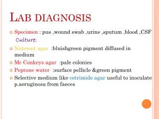
Neisseria gonorrhea Specimen: - In females: cervical swab in acute and chronic infection. • In males: urethral discharge in acute infection and morning drops in chronic infection. • Other specimens that may be used: i- throat swab. ii- anorectal swab iii- conjunctival swab in case of neonatal conjunctivitis. 1- gram stain: gm-ve kidney-shaped diplococci that are seen extracellularly and intracellularly inside PNL. 2- Culture on Thayer-Marten agar (chocolate agar that contains vancomycin, nystatin, colistin and trimethoprim) to suppress normal flora..
Why chocolate agar and not blood agar? Blood agar contains substances which inhibit the growth of Neisseria as trace metals and fatty acids. Heating blood to 80°C (chocolate agar) inactivates these inhibitors. - Incubation conditions: aerobically at 36ºC for 24-48 hours in presence of 5% CO2. Suspected colonies are identified by: a- morphology of the colonies on the culture plates. b- gram stain from the colonies c- BR:
- oxidase test: Oxidase +ve (i.e. produce cytochrome oxidase) which is demonstrated by using filter paper impregnated with a dye (tetramethyl phenylenediamin) that on oxidation changes color to dark purple. - Sugar fermentation: Ferment glucose only. Does not ferment maltose or sucrose. 3- Gonococcal antigens in the specimen can be detected by ELISA. 4- DNA probe to detect gonococcal ribosomal genes. Serological diagnosis: - detection of specific antibodies to gonococcal pilin and outer proteins by ELISA.
Gm-stained smear of pathological discharge of case of gonorrhea showing gm-ve diplococci inside & outside pus cells (PNLs)
Morphology of N. gonorrhea on modified chocolate agar (Thayer-Marten agar)
Gm stain from colonies of N. gonorrhea Gm-ve kidney-shaped diplococci
Oxidase test Sugar fermentation can be used to differentiate pathogenic from commensalneisseria.
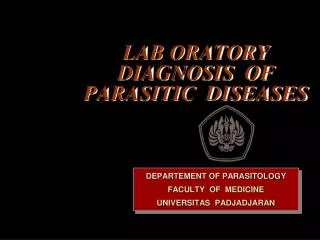
Treponema pallidum Syphilis I- Detection of T. pallidum in lesions: Serous exudate from lesions of 1ry and 2ry stages is examined by: 1- wet film for dark ground microscopy to detect the darting movement of the spiral T. pallidum. 2- IFT using fluorescein-labelled antitreponemal antibodies. 3- staining with silver impregnation technique (Fontana stain).
Dark-ground microscopy showing long slender spiral bacteria Treponema pallidum IF staining showingthe spiral Treponema pallidum
Treponema pallidum smear Stained by silver impregnation Technique (Fontana Stain) showing the spiral morphology
II- Serological diagnosis: A- non-treponemal antigen tests: • detect the reagin antibody that react with a non-specific antigen, cardiolipin, which is an alcohol extract of beef heart muscle supplemented with lecithin and cholesterol. • Reagin is a mixture of IgG and IgM that appear 2-3 weeks in the patient serum and 4-8 weeks in the CSF after exposure to infection. 1-Flocculation tests: VDRL and RPR (rapid plasma reagin) tests. 2- CFT, complement fixation test (Wasserman test).
non-treponemal antigen tests are characterized by: - Non-specific and can lead to false positive results. - Become negative 6-18 months after effective treatment, so can be used to follow up the effect of treatment. - They are mainly used for screening and epidemiological studies because they are sheep, rapid and simple.
The rapid plasma reagin (RPR) test - The upper, left well shows a negative test result, the carbon particles have remained unclumped. - The clumped appearance of the carbon particles in the right well indicate a positive test result, illustrating the flocculation of the cardiolipin-based antigen by antibodies in the test serum.
B- Treponemal antigen tests - Highly specific and sensitive tests as they use T. pallidum as the antigen. - But they are complex and expensive, so used mainly for confirmation of diagnosis. -They remain positive for life, so cannot be used to judge the efficacy of treatment. 1- Fluorescent treponema antibody test (FTA). The presence of IgM FTA in the blood of a new-borne is good evidence of in-utero infection. 2-Treponema pallidum hemagglutination (TPHA). 3- T. pallidum Particle Agglutination (TPPA).
1- Fluorescent treponema antibody test (FTA). 2- TPHA
Lymphogranuloma venerium Chlamydia trachomatis serotypes L1-L3 Specimen: Smears from the lesion and aspirate from the LN 1- stained by Giemsa or IF to see the intracytoplasmic inclusions. 2- Direct detection of chlamydial antigens in the specimen using specific monoclonal antibodies by ELISA or IFT. 3- Direct detection of nucleic acid by PCR and DNA probes. 4- Isolation in tissue culture. Serological diagnosis to detect specific IgM or high titer of IgG by ELISA or IFT.
Chlamydia trachomatis serotypes D-K Non-gonococcalurethritis Specimen: - In female: endocervical swab - In male: urethral dicharge. - Conjunctival swab in case of neonatal conjunctivitis. Procedures: As in case of LGV
Hemophilusducreyi Chancroid Specimen: Scraping from the lesion or aspirate from the enlarged lymph nodes:- a- gram stained smear: Gm-vecoccobacilli b- culture on chocolate agar. c- Subculture on nutrient agar containing 2 discs for X and V factors. Growth is enhanced around X factor only. 2- Serological diagnosis
Gm-ve coccobacilli Ex: H. ducreyi Growth apprears around X factor No growth around V factor Growth on chocolate agar
Mycoplasma hominis Specimen: • female: endocervical swab • Male: urethral discharge 1-Culture of the specimen on specific media→ fried egg appearance. 2- Direct detection of Mycoplasma antigen in the specimens by IFT and PCR. 3- Serological diagnosis: detection of specific IgM or rising titer of IgG in the patient serum.
Fried egg appearance of mycoplasma colonies on the specific medium
Candida vulvovaginitis Mycotic vulvovaginitis Specimen: Swab from the curd like white vaginal discharge 1- gram stain many epithelial cells and strongly gm+ve budding yeast cells and pseudohypha. 2- culture on Sabouraud dextrose agar Large white colonies.
Gm stain of vaginal discharge In case of candida vaginitis→ Many epithelial cells and gm+ve budding candida yeast and pseudohyphae Colonies of Candida albicans on Sabouraud dextrose agar