Download

1 / 19
200 likes | 296 Vues
Learn how the body regulates acid-base balance, compensates for derangements, and identifies metabolic acidosis causes and treatments.

E N D
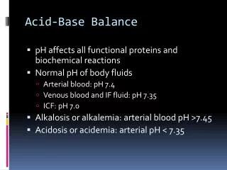
Acid-Base Balance Acid-Base Homeostasis The pH of body fluids is maintained within a narrow range despite the ability of the kidneys to generate large amounts of HCO3– and the normal large acid load produced as a by-product of metabolism. This endogenous acid load is efficiently neutralized by buffer systems and ultimately excreted by the lungs and kidneys.
Important buffers include intracellular proteins and phosphates and the extracellular bicarbonate–carbonic acid system. Compensation for acid-base derangements can be by respiratory mechanisms (for metabolic derangements) or metabolic mechanisms (for respiratory derangements). Changes in ventilation in response to metabolic abnormalities are mediated by hydrogen-sensitive chemoreceptors found in the carotid body and brain stem. Acidosis stimulates the chemoreceptors to increase ventilation, whereas alkalosis decreases the activity of the chemoreceptors and thus decreases ventilation.
The kidneys provide compensation for respiratory abnormalities by either increasing or decreasing bicarbonate reabsorption in response to respiratory acidosis or alkalosis, respectively. Unlike the prompt change in ventilation that occurs with metabolic abnormalities, the compensatory response in the kidneys to respiratory abnormalities is delayed. Significant compensation may not begin for 6 hours and then may continue for several days. Because of this delayed compensatory response, respiratory acid-base derangements before renal compensation are classified as acute, whereas those persisting after renal compensation are categorized as chronic.
The predicted compensatory changes in response to metabolic or respiratory derangements are listed in Table 3-7.18 If the predicted change in pH is exceeded, then a mixed acid-base abnormality may be present (Table 3-8). • derangements are listed in Table 3-7.18 If the predicted change in pH is exceeded, then a mixed acid-base abnormality may be present
Metabolic Derangements Metabolic Acidosis Metabolic acidosis results from an increased intake of acids, an increased generation of acids, or an increased loss of bicarbonate (Table 3-9). The body responds by several mechanisms, including producing buffers (extracellular bicarbonate and intracellular buffers from bone and muscle), increasing ventilation (Kussmaul's respirations), and increasing renal reabsorption and generation of bicarbonate. The kidney also will increase secretion of hydrogen and thus increase urinary excretion of NH4+ (H+ + NH3+ = NH4+). Evaluation of a patient with a low serum bicarbonate level and metabolic acidosis includes determination of the anion gap (AG), an index of unmeasured anions.
The normal AG is <12 mmol/L and is due primarily to the albumin effect, so that the estimated AG must be adjusted for albumin (hypoalbuminemia reduces the AG).19 Metabolic acidosis with an increased AG occurs either from ingestion of exogenous acid such as from ethylene glycol, salicylates, or methanol, or from increased endogenous acid production of the following: -Hydroxybutyrate and acetoacetate in ketoacidosis Lactate in lactic acidosis Organic acids in renal insufficiency
A common cause of severe metabolic acidosis in surgical patients is lactic acidosis. In circulatory shock, lactate is produced in the presence of hypoxia from inadequate tissue perfusion. The treatment is to restore perfusion with volume resuscitation rather than to attempt to correct the abnormality with exogenous bicarbonate. With adequate perfusion, the lactic acid is rapidly metabolized by the liver and the pH level returns to normal. The administration of bicarbonate for the treatment of metabolic acidosis is controversial, because it is not clear that acidosis is deleterious.20 The overzealous administration of bicarbonate can lead to metabolic alkalosis, which shifts the oxyhemoglobin dissociation curve to the left; this interferes with oxygen unloading at the tissue level and can be associated with arrhythmias that are difficult to treat. An additional disadvantage is that sodium bicarbonate actually can exacerbate intracellular acidosis
Administered bicarbonate can combine with the excess hydrogen ions to form carbonic acid; this is then converted to CO2 and water, which thus raises the partial pressure of CO2 (PCO2). This hypercarbia could compound ventilation abnormalities in patients with underlying acute respiratory distress syndrome. This CO2 can diffuse into cells, but bicarbonate remains extracellular, which thus worsens intracellular acidosis. Clinically, lactate levels may not be useful in directing resuscitation, although lactate levels may be higher in nonsurvivors of serious injury.21
Metabolic acidosis with a normal AG results either from exogenous acid administration (HCl or NH4+), from loss of bicarbonate due to GI disorders such as diarrhea and fistulas or ureterosigmoidostomy, or from renal losses. In these settings, the bicarbonate loss is accompanied by a gain of chloride; thus, the AG remains unchanged. To determine if the loss of bicarbonate has a renal cause, the urinary [NH4+] can be measured. A low urinary [NH4+] in the face of hyperchloremic acidosis would indicate that the kidney is the site of loss, and evaluation for renal tubular acidosis should be undertaken. Proximal renal tubular acidosis results from decreased tubular reabsorption of HCO3–, whereas distal renal tubular acidosis results from decreased acid excretion. The carbonic anhydrase inhibitor acetazolamide also causes bicarbonate loss from the kidneys.
Metabolic Alkalosis Normal acid-base homeostasis prevents metabolic alkalosis from developing unless both an increase in bicarbonate generation and impaired renal excretion of bicarbonate occur (Table 3-10). Metabolic alkalosis results from the loss of fixed acids or the gain of bicarbonate and is worsened by potassium depletion. The majority of patients also will have hypokalemia, because extracellular potassium ions exchange with intracellular hydrogen ions and allow the hydrogen ions to buffer excess HCO3–. Hypochloremic, hypokalemic, and metabolic alkalosis can occur from isolated loss of gastric contents in infants with pyloric stenosis or adults with duodenal ulcer disease.
Unlike vomiting associated with an open pylorus, which involves a loss of gastric as well as pancreatic, biliary, and intestinal secretions, vomiting with an obstructed pylorus results only in the loss of gastric fluid, which is high in chloride and hydrogen, and therefore results in a hypochloremic alkalosis. Initially the urinary bicarbonate level is high in compensation for the alkalosis. Hydrogen ion reabsorption also ensues, with an accompanied potassium ion excretion. In response to the associated volume deficit, aldosterone-mediated sodium reabsorption increases potassium excretion. The resulting hypokalemia leads to the excretion of hydrogen ions in the face of alkalosis, a paradoxicaciduria. Treatment includes replacement of the volume deficit with isotonic saline and then potassium replacement once adequate urine output is achieved.
Respiratory Derangements Under normal circumstances blood PCO2 is tightly maintained by alveolar ventilation, controlled by the respiratory centers in the pons and medulla oblongata. Respiratory Acidosis Respiratory acidosis is associated with the retention of CO2 secondary to decreased alveolar ventilation. The principal causes are listed in Table 3-11. Because compensation is primarily a renal mechanism, it is a delayed response. Treatment of acute respiratory acidosis is directed at the underlying cause.
Measures to ensure adequate ventilation are also initiated. This may entail patient-initiated volume expansion using noninvasive bilevel positive airway pressure or may require endotracheal intubation to increase minute ventilation. In the chronic form of respiratory acidosis, the partial pressure of arterial CO2 remains elevated and the bicarbonate concentration rises slowly as renal compensation occurs.
Respiratory Alkalosis In the surgical patient, most cases of respiratory alkalosis are acute and secondary to alveolar hyperventilation. Causes include pain, anxiety, and neurologic disorders, including central nervous system injury and assisted ventilation. Drugs such as salicylates, fever, gram-negative bacteremia, thyrotoxicosis, and hypoxemia are other possibilities. Acute hypocapnia can cause an uptake of potassium and phosphate into cells and increased binding of calcium to albumin, leading to symptomatic hypokalemia, hypophosphatemia, and hypocalcemia with subsequent arrhythmias, paresthesias, muscle cramps, and seizures. Treatment should be directed at the underlying cause, but direct treatment of the hyperventilation using controlled ventilation may also be required.