Download
1 / 66
670 likes | 1.02k Vues
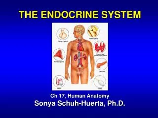
THE ENDOCRINE SYSTEM Ch 17, Human Anatomy Sonya Schuh-Huerta, Ph.D. The Endocrine System: An Overview. A system of ductless glands Secrete messenger molecules called hormones Interacts closely with the nervous system Endocrinology Study of hormones & endocrine glands. Endocrine Organs.

E N D
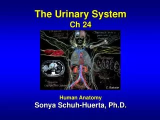
THE ENDOCRINE SYSTEM Ch 17, Human Anatomy Sonya Schuh-Huerta, Ph.D.
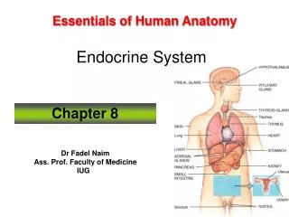
The Endocrine System: An Overview • A system of ductless glands • Secrete messenger molecules called hormones • Interacts closely with the nervous system • Endocrinology • Study of hormones & endocrine glands
Endocrine Organs • Scattered throughout the body • Pure endocrine organs • Pituitary, pineal, thyroid, parathyroid, & adrenal glands • Organs containing endocrine cells • Pancreas, thymus, gonads, & hypothalamus • Hypothalamus is a neuroendocrineorgan • Produces hormones & also has nervous functions
Location of the Major Endocrine Glands Pineal gland Hypothalamus Pituitary gland Thyroid gland Parathyroid glands (on dorsal aspect of thyroid gland) Thymus Adrenal glands Pancreas Ovary (female) Testis (male)
Hormones • Classes of hormones • Amino acid–based hormones • Steroids derived from cholesterol • Basic hormone action • Circulate throughout the body in the blood • Influences only specific tissues target cells • A hormone can have different effects on different target cells
Control of Hormone Secretion • Secretion triggered by 3 major types of stimuli • Humoral simplest of endocrine control mechanisms • Secretion in direct response to changing ion or nutrient levels in the blood • Parathyroid monitors calcium levels • Responds to decline by secreting hormone to reverse decline
Control of Hormone Secretion • Secretion triggered by 3 major types of stimuli (cont…) • Neural • Nerve fibers stimulate hormone secretion • Sympathetic nerve fibers stimulate cells in the adrenal medulla • Induces release of epinephrine & norepinephrine • Hormonal • Certain hormones trigger secretion of other hormones • Hypothalamus secretes hormones stimulates pituitary to secrete hormones stimulates other glands to secrete hormones
(a) Humoral stimulus (b) Neural stimulus (c) Hormonal stimulus 1 1 1 Capillary blood contains low concentration of Ca2+, which stimulates… Preganglionic sympathetic fibers stimulate adrenal medulla cells… The hypothalamus secretes hormones that… Hypothalamus CNS (spinal cord) Capillary (low Ca2+ in blood) 2 …stimulate the anterior pituitary gland to secrete hormones that… Thyroid gland (posterior view) Parathyroid glands Pituitary gland Preganglionic sympathetic fibers Adrenal cortex Thyroid gland Gonad (Testis) Medulla of adrenal gland Parathyroid glands PTH Capillary 3 2 2 …secretion of parathyroid hormone (PTH) by parathyroid glands. PTH acts to increase blood Ca2+. …to secrete catecholamines (epinephrine and norepinephrine) …stimulate other endocrine glands to secrete hormones 3 Major Types of Stimuli
Control of Hormone Secretion • Always controlled by feedback loops • Blood concentration declines below a minimum • More hormone is secreted • Blood concentration exceeds maximum • Hormone production is halted • Referred to as positive & negativefeedback loops usually multiple hormones involved in the pathway
The Pituitary Gland • Secretes 9 major hormones • Attached to the hypothalamus by the infundibulum (= pituitary stalk) • 2 basic divisions of the pituitary gland: • Adenohypophysis (= anterior pituitary) • Has 3 major divisions • Neurohypophysis (= posterior pituitary) has 2 major divisions
Corpus callosum Thalamus Pineal Acidophil Basophil Chromophobe cell Hypothalamus Mammillary body Brain stem Pituitary (hypophysis) (b) Capillary with red blood cells Spherical cluster of cells (a) (d) The Pituitary Gland Mammillary body Optic chiasma Median eminence of hypothalamus Tuber cinereum Anterior lobe Posterior lobe Pars tuberalis Pars intermedia Infundibulum Pars distalis Pars nervosa (c)
Anterior Pituitary • The largest division of the anterior lobe: • Makes & secretes 7 different hormones • Tropic hormones regulate the hormone secretion of other glands • Includes: TSH, ACTH, FSH, LH
Anterior Pituitary • GH, PRL, & MSH • Act directly on non-endocrine target tissues
Anterior Pituitary • Growth hormone/GH (= somatotropic hormone) • Produced by somatotropic cells • Stimulates body growth by stimulating increased protein production & growth of epiphyseal plates • Stimulates growth directly & indirectly by the liver’s secretion of insulin-like growth factor-1 (IGF1)
Anterior Pituitary • Thyroid-stimulating hormone (TSH) • Produced by thyrotropic cells • Signals thyroid gland to secrete thyroid hormones • Adrenocorticotropic hormone (ACTH) • Stimulates adrenal cortex to secrete hormones that help cope with stress
Anterior Pituitary • Melanocyte-stimulating hormone (MSH) • In humans, MSH functions in appetite supression (“My Stomach’s not Hungry” hormone) • Gonadotropins FSH & LH • – Produced by gonadotropic cells • – they increase sex steroid production by the • gonads & the maturation of gametes
Anterior Pituitary • Prolactin (PRL) produced by prolactin cells • Targets milk-producing glands in the breast stimulates milk production after childbirth & • during lactation • There are prolactin-secreting tumors that result in milk production in both women & men • (galactorrhea; can lead to problems with fertility) • Prolactin central to milk production in all female mammals (exception – male bats)
Hypothalamic Control of Hormone Secretion from the Anterior Lobe • The hypothalamus • Controls secretion of anterior lobe hormones • Exerts control by secreting: • Releasing hormones prompt anterior lobe to release hormones • Gonadotropin-releasing hormone (GnRH) produced & secreted by specific neurons of hypothalamus cause release of FSH & LH • Inhibiting hormones turn off secretion of anterior lobe hormones
Hypothalamic Control of Hormone Secretion from the Anterior Lobe • Releasing hormones • Are secreted like neurotransmitters • Enter a primary capillary plexus • Travel in hypophyseal portal veins to a secondary capillary plexus • From the secondary capillary plexus, the releasing hormones trigger the anterior lobe to secrete the specific hormone, which is dumped into the secondary capillary plexus, and enters general circulation & travels to target organs
When appropriately stimulated, hypothalamic neurons secrete releasing and inhibiting hormones into the primary capillary plexus. 1 Hypothalamic neuron cell bodies Hypothalamus Superior hypophyseal artery Hypophyseal portal system Primary capillary plexus Hypothalamic hormones travel through the portal veins to the anterior pituitary where they stimulate or inhibit release of hormones from the anterior lobe. 2 Hypophyseal portal veins Secondary capillary plexus Anterior lobe of pituitary Anterior pituitary hormones are secreted into the secondary capillary plexus. 3 TSH, FSH, LH, ACTH, GH, PRL (a) Relationship between the anterior pituitary & hypothalamus Hypothalamic Control of Ant. Pituitary Secretion
Posterior Pituitary • Is structurally part of the brain • Its axons make up the hypothalamic–hypophyseal tract • Arises from neuronal cell bodies in the hypothalamus • Supraoptic nucleus • Paraventricular nucleus
Relationship Between Posterior Pituitary & Hypothalamus Hypothalamic neurons synthesize oxytocin and ADH. 1 Hypothalamus Paraventricular nucleus Supraoptic nucleus Oxytocin and ADH are transported along the hypothalamic- hypophyseal tract to the posterior lobe. 2 Optic chiasma Infundibulum (connecting stalk) Inferior hypophyseal artery Hypothalamic- hypophyseal tract 3 Oxytocin and ADH are stored in axon terminals in the posterior pituitary. Axon terminals Oxytocin and ADH are released into the blood when hypothalamic neurons fire. Posterior lobe of pituitary 4 Oxytocin ADH (b) Relationship between the posterior pituitary & hypothalamus
Posterior Pituitary • Does not make hormones • Stores & releases hormones made in the hypothalamus! • Releases 2 very important peptide hormones: • Anti-Diuretic hormone (ADH) –remember this? • Oxytocin
Posterior Pituitary • ADH (Anti-Diuretic hormone; =vasopressin) • Targets kidneys to resorb water; prevents diuresis • Oxytocin • Induces smooth muscle contraction of reproductive organs, ejects milk during breast feeding, & signals contraction of the uterus during childbirth
Hormones Made by Hypothalamus & Secreted by Posterior Pituitary
Pineal Gland • Located on the roof of the diencephalon • Shaped like a pine cone • Pinealocytes secrete melatonin • A hormone that regulates circadian rhythms
Thyroid Gland • Located in anterior neck around trachea • Largest pure endocrine gland • Functions in metabolism & metabolic rate; how quickly the body uses energy; making proteins, etc. • Composed of follicles & areolar connective tissue
Hyoid bone Epiglottis Thyroid cartilage External carotid artery Superior thyroid artery Common carotid artery Inferior thyroid artery Isthmus of thyroid gland Right subclavian artery Left subclavian artery Left lateral lobe of thyroid gland Trachea Aorta (a) Gross anatomy of the thyroid gland, anterior view Thyroid Gland
Thyroid Gland Hormones • Produces 2 types of hormones: • Thyroid hormones (TH = T3 & T4) • Thyroxine (T4) & triiodothyronine (T3) tyrosine-based hormones produced by thyroid • Involved in regulation of metabolism act on nearly all cells of body; increase basal metabolic rate, protein synthesis; long bone growth; increase body’s sensitivity to catecholamines; regulate protein, fat & carb metabolism • Secreted by follicular cells • Calcitonin Secreted by parafollicular cells; lowers blood Ca2+ levels (inhibits intestinal Ca2+ absorption & inhibits osteoclast activity in bones; protects against calcium loss from skeleton during pregnancy & lactation
Microscopic Anatomy of Thyroid Gland Colloid-filled follicles Follicular cells (secrete thyroid hormone) Parafollicular cell (secretes calcitonin) (b) Photomicrograph of thyroid gland follicles (160)
Parathyroid Glands • Lie on the posterior surface of thyroid gland • Contain 2 types of endocrine cells • Chief cells • Produce Parathyroid Hormone (PTH) • Functions to increase blood Ca2+ levels by acting on receptors in bone, kidney & intestine all leads to higher plasma calcium levels! (opposite of calcitonin) • Oxyphil cells • Function unknown...
Parathyroid Glands: Gross & Microscopic Pharynx (posterior aspect) Parathyroid cells (secrete parathyroid hormone) Thyroid gland Parathyroid glands Esophagus Oxyphil cells Trachea Capillary (a) Location of parathyroid glands, posterior view (b) Photomicrograph of parathyroid gland tissue (360)
Adrenal Glands (Suprarenal Glands) • Pyramid-shaped glands located on the superior surface of each kidney • Supplied by about 60 suprarenal arteries • Nerve supply is almost exclusively sympathetic fibers!!! • Functions in…? -remember?
Adrenal Glands • 2 endocrine glands in one! • Adrenal medulla a cluster of neurons • Part of Sympathetic Nervous System! • Postganglionic nerve fibers • Adrenal cortex forms the bulk of the gland • Functions All adrenal hormones help one cope with danger, terror, or stress
Adrenal Medulla • Chromaffin cells • Are modified ganglionic sympathetic neurons • Secrete amine hormones epinephrine & norepinephrine (= catecholamines) • Enhance “fight-or-flight” response • Hormones are stored in secretory vesicles • Are arranged in spherical clusters & some branching cords
Adrenal Cortex • Secretes lipid-based steroid hormones glucocorticoids, mineralocorticoids, androgens, E2 • Cortex is composed of 3 layers (zones): • Zona glomerulosa cells arranged in spherical clusters • Zona fasciculata cells arranged in parallel cords; contains lipid droplets • Zona reticularis cells arranged in a branching network
The Adrenal Gland: Gross & Microscopic Hormones secreted Capsule Zona glomerulosa Aldosterone Zona fasciculata Adrenal gland Cortex Cortisol and androgens Medulla Cortex Zona reticularis Kidney Adrenal medulla Medulla Epinephrine and norepinephrine (a)Drawing of the histology of the adrenal cortex and a portion of the adrenal medulla (b) Photomicrograph (140X)
Short-term stress More prolonged stress Stress Nerve impulses Hypothalamus CRH (corticotropin- releasing hormone) Spinal cord Corticotroph cells of anterior pituitary Preganglionic sympathetic fibers To target in blood Adrenal cortex (secretes steroid hormones) Adrenal medulla (secretes amino acid– based hormones) ACTH Catecholamines (epinephrine and norepinephrine) Mineralocorticoids Glucocorticoids Short-term stress response Long-term stress response 1.Increased heart rate 2.Increased blood pressure 3.Liver converts glycogen to glucose and releases glucose to blood 4.Dilation of bronchioles 5.Changes in blood flow patterns leading to decreased digestive system activity and reduced urine output 6.Increased metabolic rate 1.Retention of sodium and water by kidneys 2.Increased blood volume and blood pressure 1.Proteins and fats converted to glucose or broken down for energy 2.Increased blood glucose 3.Suppression of immune system Stress & the Adrenal Gland
Pancreas • Located in the posterior abdominal wall • Functions regulation of blood sugar, metabolism & digestion • Contains endocrine & exocrine cells • Exocrine cells • Acinar cells secrete digestive enzymes (remember?..) • Endocrine cells • Islets of Langerhans • About one million islets scattered throughout the pancreas
Islets of Langerhans • Main endocrine cell types: • Alpha cells ( cells) secrete glucagon • Signal liver to release glucose from glycogen • Raises blood sugar • Beta cells ( cells) secrete insulin • Signal most body cells to take up glucose from the blood • Promotes storage of glucose as glycogen in liver • Lowers blood sugar
Pancreas • Pancreatic islets contain 2 rare cell types • Delta (∂) cells • Secrete somatostatin • Inhibits secretion of insulin & glucagon • F (PP) cells • Secrete pancreatic polypeptide (PP) • May inhibit exocrine activity of the pancreas
Thymus • Located in the lower neck & anterior thorax • Function Important immune organ; T-lymphocytes arise from precursor cells here; decreases in size & function after puberty
Gonads • Main sources of sex steroid hormones • Testes & ovaries • Male • Interstitial Leydigcells secrete androgens • Primarily testosterone • Promotes development of the testes • Promotes the formation & survival of sperm • Development & maintenance of secondary sex characteristics
Gonads • Female • Ovaries • Androgens & estrogens secreted by follicular cells • Estrogen • Dev & maintenance of secondary sex • characteristics • – Dev of eggs • Regulation of menstrual cycle • Progesterone • Prepares the uterus for Pregnancy
Sex Steroid Hormones • Critical for normal reproductive function • Affect many other tissues/organs of the body • Bone • Muscle • Fat • Liver & kidney • Brain (sexual behavior, mood, cognition)
2008 2010 Sex Steroid Hormones & Bone • Promote bone growth (adolescent growth spurt)
Sex Steroid Hormones & Bone • Also maintain bone mass • With aging or in diseases that render the gonads dysfunctional (low T/E2) loss of bone mass & osteoporosis!
Could Skeletal Tissue Exert Reciprocal Effects on Gonads? ??? sex steroids gonad bone