Download
1 / 21
230 likes | 383 Vues
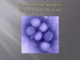
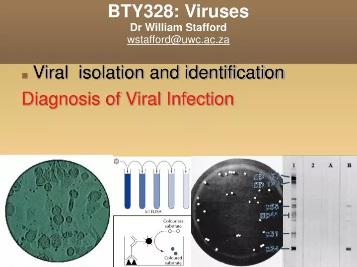
BTY328: Viruses Dr William Stafford wstafford@uwc.ac.za. Viral isolation and identification Diagnosis of Viral Infection. Overview of methods to identify virus. Histological and cellular changes (Cytopathic effects, haemadsorption) Formation of plaques Serological methods (IF, ELISA)

E N D
BTY328: VirusesDr William Staffordwstafford@uwc.ac.za • Viral isolation and identification Diagnosis of Viral Infection
Overview of methods to identify virus • Histological and cellular changes(Cytopathic effects, haemadsorption) • Formation of plaques • Serological methods (IF, ELISA) • Electron microscopy • DNA molecular methods (PCR, hybridisation)
Cytopathic Effect Cytopathic effect of enterovirus 71 and HSV in cell culture: note the ballooning of cells. (Virology Laboratory, Yale-New Haven Hospital, Linda Stannard, University of Cape Town)
Haemadsorption Syncytial formation caused by mumps virus and haemadsorption of erythrocytes onto the surface of the cell sheet. (courtesy of Linda Stannard, University of Cape Town, S.A.)
1:100 1:10 1:10 1:10 1:10 1:10 virus serial dilution 10-2 10-3 10-4 10-5 10-6 10-7 plate 1 ml plaques (100,000) (10,000) (1000) 100 10 1 Titer = 1 x 10⁷ pfu/ml Plaque assay: method
Plaque assay Serial dilution to find viral titre: With and without (+/-) IBT antiviral drug Fields Virology, 4th ed, Knipe & Howley, eds, Lippincott Williams & Wilkins, 2001, Fig. 2-5
ELISA: HIV detection Microplate ELISA for HIV antibody: colored wells indicate reactivity
Western Blot HIV-1 Western Blot • Lane1: Positive Control • Lane 2: Negative Control • Sample A: Negative • Sample B: Indeterminate • Sample C: Positive
Hemagglutination assay 1:8 1:2 1:2 1:2 1:2 1:2 virus serial dilution 8 16 32 64 128 256 mix with red blood cells side view top view Titer = 32 HA units/ml
Haemagglutination assay From Medical Microbiology, 5th ed., Murray, Rosenthal & Pfaller, Mosby Inc., 2005, Fig. 51-6.
Hemagglutination assay: influenza virus Hemagglutination assay. Seven different samples of influenza virus, numbered 1 through 7 at the left, were serially diluted as indicated at the top, mixed with chicken red blood cells (RBC), and incubated on ice for 1 to 2 hours. Wells in the bottom row contain no virus. Agglutinated RBCs coat wells evenly, in contrast to nonagglutinated cells, which form a distinct button at the bottom of the well. The HA titer, shown at the right, is the last dilution that shows complete hemagglutination activity. (From Fields Virology, 4th ed, Knipe & Howley, eds, Lippincott Williams & Wilkins, 2001, Fig. 2-8)
Immunofluorescense HSV-infected epithelial cells from skin lesion. (Source: Virology Laboratory, Yale-New Haven Hospital) Positive immunofluorescence test for rabies virus antigen. (Source: CDC)
Immune Electron Microscopy • Either transmission or scanning electro microscopy is carried to quantify viruses in a sample and determine viral structure. • EM can be enhance by using antibodies specific for a viral antigen (the antibody is usually labelled by conjugation to gold particles)
Electronmicrographs Adenovirus Rotavirus (courtesy of Linda Stannard, University of Cape Town, S.A.) |____________________| Approx. 100nm
Direct particle count using electron microscopy Direct electron microscopic particle count. An electron micrograph of a spray droplet containing 15 latex beads (spheres) and 14 vaccinia virus particles (slightly smaller, brick-shaped particles). (From Fields Virology, 4th ed, Knipe & Howley, eds, Lippincott Williams & Wilkins, 2001, Fig. 2-7.)
DNA Molecular Techniques • Dot-blot, Southern blot, in-situ hydridization are examples of classical techniques. depend on the use of specific DNA/RNA probes for hybridization. • PCR for specific viral genes • Whole viral genome sequencing.
Viral isolation: Differential centrifugation • Partial purification may be achieved by resistance to chemicals (CHCl3), enzymes (DNase, Rnase). • Viruses can also be • separated from host cells • by size selected filtrationand differential centriguation • (10 000g and 10000 g).
Virus isolation by density gradient centrifugation Equilibrium density gradient centrifugation and rate zonal centrifugation • Equilibrium density gradient centrifugation • and Rate zonal centrifugation separates viruses from cells and cellular components based on their size and density. • Purification of specific viruses can be achieved by affinity chromatography using antibodies directed to the virus of interest.
Summary: Virus identification and isolation • Main clinical diagnostic techniques • Cell culture, serology and antigen detection, nucleic acid detection • Virus culture • Cytopathic effect • Not all viruses can be cultured! • Virus quantitation • Biological in vivo methods (palque assay and LD50/ID50 for animal models) • Physical (serological assays, heamagglutination, electron microscopy) • Isolation of viruses and infectious agents by physico-chemical methods • Nature and identification of viruses, (also prions, viroids…!?)