Lupus nephritis
550 likes | 779 Vues
Lupus nephritis. review. Introduction. Tissue injury can affect nearly everyorgan, however, it is the damage to the kidney that is largely responsible for morbidity and mortality Within the kidney the expression of SLE is heterogeneous.

Lupus nephritis
E N D
Presentation Transcript
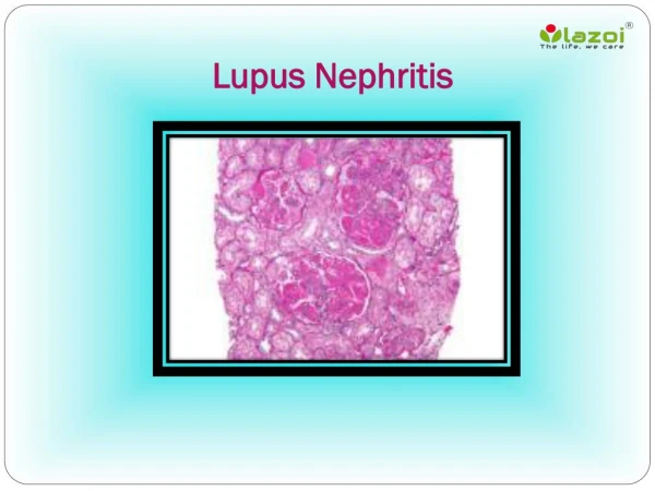
Lupus nephritis review
Introduction • Tissue injury can affect nearly everyorgan, however, it is the damage to the kidney that is largely responsible for morbidity and mortality • Within the kidney the expression of SLE is heterogeneous.
it is particularly pressing that we dissect the genetics and pathogenesis of this complex disease that targets the kidney • it is not surprising that there has not been a new therapeutic approved to treat patient’s with SLE for more than 40 years.
Genetics of lupus • The inheritance of this type of diseases has been coined threshold liability • a threshold liability disease develops when an individual accumulates a number of • genetic and environmental factors that is greater than the disease threshold.
The involvement of nongenetic factors in SLE susceptibility is shown clearly by a concordance rate between monozygotic twins of 34% to 50% • Genetic analyses of inbred murine models of systemic autoimmunity also strongly argue in favor of a genetic basis for SLE susceptibility.
The exact number of genes involved in SLE susceptibility currently is unknown, an • it is most likely that SLE susceptibility results from various combinations of different genes. • With a few exceptions, the identity of these genes also is unknown.
Association studies evaluate candidate genes that have been selected based on their function in the immune system (eg, Fc fragment of IgG, low affinity IIIa, receptor [FCGRIIIA]), • or their aberrant expression in lupus patients (eg, interleukin [IL]-8)
Linkage analyses rely on genome-wide scans of families of SLE patients with anonymous DNA markers. • Subsequent statistical analyses have mapped the location of genomic regions • named quantitative trait loci (QTL)-linked SLE or LN susceptibility
IS THERE A GENETICPREDISPOSITION TO LN? • A number of studies, favor a genetic basis for renal involvement in SLE. • The risk of developing end-stage renal disease is 2.6- to 5.6-fold greater in African Americans than in European Americans • a number of studies have shown a genetic component to end-stage renal disease
Because LN is one of the most serious clinical outcomes of SLE, it is possible, however, that • renal involvement represents a marker of disease severity rather than a distinct genetic cause.
LN also is associated strongly with a number of other disease markers eg anti dsDNA. • It is therefore possible that some of the LN susceptibility genes are in fact genes that predispose to the production of these antibodies.
there is evidence that LN genetic basis is supported both by genes that • target the pathogenic autoimmune effector response to the kidneys • and by genes that increase the global severity of the autoimmune • pathology, and therefore increase the likelihood • of renal disease in these patients.
Genes That Increase Kidney Susceptibility to Autoimmune Pathogenesis • Among the genes that have been associated with LN, angiotensin-converting enzyme (ACE) and angiotensinogen (AGT), • 2 essential genes from the renin angiotensin system, are the best illustration of the existence of renal-specific factors.
LN is a prototypic immune complex–mediated disease and it has been proposed that the • low-affinity IgG receptor FCRIIIA (CD16) • plays an essential role in immune-complex clearance, preventing the deposition of pathogenic autoantibodies in the kidney
Genes That AmplifyAutoimmune Pathogenesisand Increase LN Incidence or Severity • The mechanism by which genes listed in this category contribute to LN is undefined. • For most of them, however, their known function • in the regulation of the immune system suggests • that they contribute to the severity of the • systemic autoimmune pathology
For SLE, significant association has been reported with human leukocyte • antigen (HLA) class II (DRB1 and DQA) or • class III (C4 and tumor necrosis factor alpha)genes • In addition, a genome-wide analysis • has provided a strong evidence of linkage between the MHC locus and SLE.
The only other immune-related gene whose association with LN has been validated independently is IL-10.
pathogenesis A brief overview
Leukocyte–Renal Epithelial CellInteractions Regulate Lupus Nephritis • renal parenchymal cells are active participants that regulate immune responses in the kidney, • and that the interaction between parenchymal cells and leukocytes (macrophages, T cells) • determine whether the kidney is protected or destroyed during lupus nephritis
the dogma was that the kidney was • an innocent bystander, and that circulating antigen– antibody complexes lodge in the renal vascular walls, attract polymorphonuclear leukocytes, • and these leukocytes release mediators that destroy the kidney
(1) the kidney is not an innocent bystander but rather parenchymal cells are active participants that regulate immune responses in the kidney, • (2) macrophages initiate renal parenchymal cell destruction (apoptosis), and • (3) the interaction between parenchymal cells and leukocytes (macrophages,T cells) can determine whether the kidney is protected or destroyed.
MRL-Faslpr mice • the MRLFaslpr strain has a mutation in Faslpr • In the MRL-Faslpr mice there are spontaneous characteristic features that are pronounced including • a plethora of autoantibodies, lymphadenopathy, splenomegaly, and multiple tissues • that are damaged including the kidney, skin, • lungs, liver, salivary and lacrimal glands, joints, • and so forth
MACROPHAGES MEDIATE TUBULARAPOPTOSIS DURING NEPHRITIS • The macrophage is central to the pathogenesis of kidney diseases. These leukocytes are abundant in human and mouse lupus nephritis. • Activated macrophages, either by releasing molecules or via cell-to-cell contact, induce apoptosis in tubularepithelial cells (TECs).
Macrophage-mediated apoptosis is not limited to epithelial cells; macrophages initiate apoptosis of renal mesangial cells • The molecules responsible for inducing apoptosis in renal parenchymal cell types may differ.
some suggest that tumor necrosis factor- mediates mesangial cell apoptosis, others suggest that this cytokine does • not mediate TEC apoptosis. • Regardless of the mediators responsible for inducing apoptosis, it is clear that activated macrophages within the kidney during the initiation phase of the lesion are harmful.
CSF-1 • substantial evidence indicates that CSF-1 is linked to lupus nephritis in MRL-Faslpr mice. CSF-1 is a harbinger • of autoimmune-mediated lupus nephritis
What turns on renal parenchymal cells to generateCSF-1 during lupus nephritis? • What is the circulating factor in the autoimmune milieu that is responsible for inducing CSF-1 in the renal parenchymal cells?
factors in the circulation initiate a dynamic interaction between macrophages, • T cells, and renal parenchymal cells • that culminate in tissue injury during lupus nephritis.
Macrophage growth factors are increased in glomeruli of patients with lupus. • Upregulation of CSF-1 and another macrophage growth factor (granulocyte-macrophage growth factor), have been identified in glomeruli in patients • with lupus nephritis.
T cell regulatory systems • Antigen recognition alone is not sufficient for full T-cell activation. • T cells require 2 distinct signals to become fully activated. • The first signal is provided by the engagement of the TCR with the major histocompatibility complex and peptide complex on antigen-presenting cells (APCs). • The second costimulatory signal is provided by engagement of one or more TCRs with their specific ligands on APCs. • Signaling through the TCR alone in the absence of a costimulatory signal leads to prolonged T-cell unresponsiveness, anergy.
Pd 1 related apoptosis • PD-1, similar to CD28 and CTLA4, is a transmembraneprotein and belongs to the Ig superfamily. • PD-1 has an immunoreceptor tyrosine-based inhibitory motifs (ITIM) within its cytoplasmic tail. • PD-1 is expressed on activated T, B, and myeloid cells including macrophages.PD-1 • binds 2 known ligands, PD-L1 and PD-L2.
PD-L1 expression on parenchymal cells in peripheral tissues fueled the concept that the PD-1 pathway is central to preserving tolerance within target tissues
Role of Renal parenchymal cells • Kidneys in the vast majority of people remain normal. • To protect the kidney after random T-cell contact with parenchymal cells, T cells must be more readily inactivated than activated. • Bone marrow–derived macrophages, dendritic cells, and B cells are professional APCs. • Because TECs, nonprofessional APCs, comprise 80% of the renal cortex, • TECs are well positioned to turn on or turn off T-cell activation.
TECs do not express B7 molecules, the potent costimulatory signals that promote T-cell activation. • PD-L1 expression on a TEC line after stimulation with interferon- inhibits T-cell activation.
TECs stimulated by interferon- downregulate the proliferation of autoreactive T-cell clones • This suggests that TECs deliver signals that limit the expansion of autoreactive T cells in lupus nephritis
Tcell and B cell • Glomerular immunoglobulin (Ig) deposition and renal lymphocyte and macrophage infiltration lead to clinical renal involvement at some point in the clinical course of up to 75% of lupus patients
Lymphocytes and their soluble mediators participate at all levels in disease pathogenesis: initiation, perpetuation, amplification, regulation, tissue and organ destruction, • and disease relapse.
INITIATION OF NEPHRITOGENICAUTOIMMUNE RESPONSES • Lupus is a disease of failed immune tolerance to self. • The repertoire of Ig and T-cell receptors is vast and self-recognition is common, generated by gene recombinatorial and somatic events in the thymus and bone marrow • in the case of mature B cells, by somatic mutation in the peripheral lymphoid organs. • It is estimated that up to 75% of newly formed, and up to 40% of mature germinal center, B cells recognize selfantigens. This autoreactivity normally is held in check by regulatory mechanism that operate on developing lymphocytes in the central lymphoid organs and on mature cells in the spleen and lymph nodes
Nephritogenic Autoantibodiesand Antibody-Mediated Injury • Glomerular antibody deposition is a hallmark of lupus glomerulonephritis. • IgG is the predominant isotype deposited, but IgM and IgA also typically are present in human lupus nephritis, accompanied by C3, C4, and C1q.
Specificity for DNA is important for pathogenesis for a subset of lupus Ig, but multiple experimental models indicate that anti-DNA activity • is neither necessary nor sufficient to induce lupus nephritis
In situ formation of immune complexes may be a dominant mechanism of renal Ig deposition. • Direct binding of cross-reactive anti-DNA Ig to purified glomerular antigens, including -actinin and-enolase, and to cultured parenchymal cells,