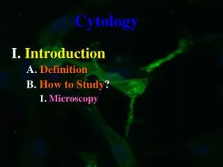
A. Definition
Cytology. I. Introduction. A. Definition. B. History. 1. Microscopes. The advantage of a microscope magnification and resolution ; Magnification to enlarge ; Resolution to clearly distinguish two objects or clarity.

A. Definition
E N D
Presentation Transcript
Cytology I. Introduction A. Definition B. History 1.Microscopes
The advantage of a microscope magnification and resolution; Magnification to enlarge; Resolution to clearly distinguish two objects or clarity a. In the 16th century, Galileo used simple pieces of glass to visualize and describe the eye of an insect. b. In the 17th century, Van Leeuwenhoekground glass to visualize the structure of cells like bacteria and sperm. c. Robert Hooke used ground glass to visualize cork structure and coined the term “cellulae” or cell.
In the 19th century Schleiden and Schwann said a. Cells are the smallest functional units of life and b. All living things are made up of cells. Later in the 19th century Virchow and Pasteur added c. Cells only arise from pre-existing cells.
II. Cytological Tools A. Microscopes 1.Light
a. Bright Field b. Dark Field c. Phase Contrast d. Confocal
a. Transmission b. Scanning c.Environmental TEM/SEM
a. Vital Stainsare mainly from variousplant pigments. forContrast
b.Antibody stains are more specific and are made byexposing antigen to some host animal. MoreContrast
III. Basic Cell Design A. Strategies 1.Prokaryotes
b. Characteristics Figure 4.4
Representative Animal Cell Figure 4.7
Representative Plant Cell Figure 4.8
B. Parts 1.Cell Membrane a. Molecular Structure
Which molecule would act as an impermeable barrier? Which molecule would act as an cellular label or antenna? Which molecule(s) would act as a transporter? Which molecule(s) would act to stiffen the membrane? Figure 4.5
Requirements= With a Concentration Gradient, Small Molecules, Requires No Energy Expenditure, and Relatively Non-polar Mechanisms= Simple Diffusion, Facilitated Diffusion, and Osmosis Page 82
Osmosis movement of a solvent (usually H2O) across a semi-permeable membrane Figure 5.13
Requirements = Uses Energy, Protein Channel, Large Molecules, and Goes against the Concentration Gradient Mechanisms = Molecular Figure 5.14
Mechanisms = Bulk If the arrowheads were reversed could you tell the difference? Figure 5.15
Mechanisms = Cell-Mediated Figure 5.16 Once inside the vesicle is the material really inside the cell?
2.Cytosol = Cell Sap a. Consistency b. Molecular Make-up
a. Cytosol consistency like thickening Jell-O b. Molecular make-up 92% is water, 7% protein, and the rest is gases, salts, lipids, and the like dissolved in the water
3.Organelles = Cell Machinery a. Membrane Bound
Nucleus = the keeper of the plans Chromatin, nucleolus envelope, and pores, Figure 4.9
Endomembrane System = rER, sER, and Golgi Figure 4.12
Energy Transformers= the Chloroplast and the Mitochondria Figure 4.15 Figure 4.14
Vacuoles = Cell storage sites Animal Types = Food (sugars, lipids, etc), or Contractile (water storage) Plant Types = Central (water storage), Amyloplasts (store starch), and Chromoplasts (store Pigments)
Cytoskeleton Figure 4.17
Ribosome and Centrioles Figure 4.19
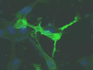
C. Cellular Specializations 1.Microvilli
Microvilli = short non-moving membrane extensions (orange area) to increase cell’s overall surface area
2.Cilia 3.Flagella
Cilia = long, moving internal cellular extensions to move something across the cell surface. Flagella = longer cellular extensions to move the entire cell Figure 4.20
Figure 4.21 i. Plants Always think function? Figure 4.11 ii. Animals Figure 4.23