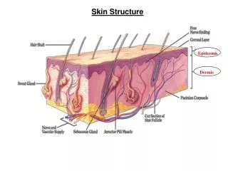
Basic Skin Structure
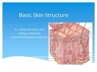
Basic Skin Structure. Dr. Salman Bin Dayel, M.D College of Medicine Salman Bin Abdulaziz University. The Skin:. The largest organ The full-thickness 1.5- to 4.0-mm Surface area about 2 square meters) It weighs about over 5KG. Functions Of The Skin:. Covers and protects the body.

Basic Skin Structure
E N D
Presentation Transcript
Basic Skin Structure Dr. Salman Bin Dayel, M.D College of Medicine Salman Bin Abdulaziz University
The Skin: • The largest organ • The full-thickness 1.5- to 4.0-mm • Surface area about 2 square meters) • It weighs about over 5KG
Functions Of The Skin: • Covers and protects the body. • Control internal temperature. • Produces vitamin D. • Receptors to detect environmental stimuli. • Regulates the movement of substances into and out of the body • Sensory function • Sociosexualcommunication • Immunological
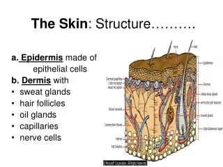
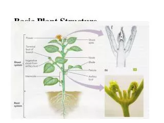
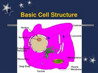
SKIN STRUCTURES: 3 Main layers: • Epidermis • The superficial portion of the skin • Dermis • The deeper layer of the skin • Primarily composed of connective tissue • Subcutaneous layer or hypodermis
EPIDERMIS • Stratified squamous epithelium • Avascular (contains no blood vessels) • 4 types of cells • 4 distinct layers of cells • Regional variation in the relative thickness • The epidermis is thickest on the palms and soles, measuring approximately 1.5 mm • It is very thin on the eyelid where it measures less than 0.1 mm
EPIDERMIS Complosed of 4 layers : • Stratum basale (germinativum) • Stratum spinosum • Stratum granulosum • Stratum corneum • In Palms and soles 5th layer called Stratum Lucidum
EPIDERMIS: Contains 4 types of cells: 1-keratinocytes (85% of cells) 2-Melanocytes 3-Langerhans cells 4-Merkel cells
Stratum basale (germinativum) • Deepest single layer of epidermis • It contains mitotically active (stem cells)10%, • columnar-shaped keratinocytes • KCs attached to each other by desmosomes & to basement membrane via keratin filaments (K5 & K14) by hemidesmosomes • Melanocyte present in this layer
Stratum spinosum: • Several cells layers • Have cytoplasmic processes (spines) • Desmosomes connect bewteen keratinocytes
Stratum granulosum: • 1 to 3 layers of fusiform shaped basophilic cells • Containes granules (Keratohyalin granules) contain profilaggrin and Loricrin proteins .
Stratum corneum: • Superifical keratinized layer Cells • Almost filled with keratin • Flattened, non nucleate • Coated with extra-cellular lipids that form water barrier of skin • Provide Mechanical protection, barrier for water loss • Layer that varies most in thickness thick palm & sole (20-30 cell layers thin other area ( 3-4 cell layers)
KERATINOCYTES: • 80-85% of epidermis • Ectoderm in origin • For keratin formation • They are arranged In many layers that continuously shed • regenerate every 4 weeks • Psoriasis 4days
MELANOCYTES: • Found in between cells of the basal layer & At the basal part of the hair follicles. • Origin neural crest • Branched cells with centeral nuclei • They produce the Melanin to keratinocytes color to the skin • No. of melanocytes for the same site is same in white and black skin. • Provide protection from UV light
LANGERHANS CELLS: • Found in upper layers of st.spinosum • Have branched shape & central nuclei • Represent 3-8%of epid. Cells • Bone marrow in origin. • Phagocytic & antigen presenting cells (Immune response)
MERKEL’S CELLS: • Found in basal cell layer • Are modified epidermal cells • Sensory nerve fibers • form terminal disk under Merkels cells • Function as touch receptors
The DERMIS: • Connective tissue layer • composed of collagen & elastic fibers, fibroblasts, macrophages & mast cells. • Contains hair follicles, glands, nerves & blood vessels • Two major regions of dermis • papillary region • reticular region
PAPILLARY DERMIS: • Upper part of dermis • Top 20% of dermis • Contain finer collagen compared to reticular dermis • Finger like projections are called dermal papillae • anchors epidermis to dermis • contains capillaries that feed epidermis • contains Meissner’s corpuscles (touch) & free nerve endings for sensations of heat, cold, pain, tickle, and itch
Located in the derml papillaeReceptor for light touch Meissner’s (Tactile) Corpuscle:
RETICULAR DERMIS: • The thicker deep layer • Dense irregular connective tissue • Contains interlacing collagen and elastic fibers • contain sweat glands , fat & hair follicles • Provides strength, extensibility & elasticity to skin • Contain many nerve receptors: • Ruffini corpuscles pressure touch. • Pacinian corpuscles vibratory pressure & touch
Basement membrane zone (BMZ) • It forms the interface between the epidermis and the dermis. • It is continuous along the epidermis and skin appendages. • Its width ranges between 50-90 nm.
It is divided into: 1- Plasma membrane and hemidesmosomes of basal keratinocytes. 2- Lamina lucida 3- Lamina densa 4- Sublamina densa fibrillar zone
Basement Membrane Zone: Main functions: 1- Attach the epidermis to the dermis. 2- Separate components of the epidermis and dermis. 3- Provide resistance against external shearing forces. 4- Maintain tissue architecture during remodeling and repair.
Subcutaneous fat tissue • the layer between the dermis and the fascia • The fat tissue acts as a cushion against external physical pressure, retain moisture . • The thickness of the subcutaneous tissue depends on the body site, age and other factors • Fat cells separated by the connective fibroid fat septum are called fat lobules.
Vascular channels and nerves: • Subcutaneous plexus • Deep • Dermal-subcutis interface • Large blood vessels • Subpapillary plexus • superficial • Parallel to epidermis