Chapter 42
Chapter 42. Circulation and Gas Exchange. Open and Closed Circulatory Systems. Open circulatory system- (insects, arthropods, and mollusks) blood bathes the organs directly. Fluid is called hemolymph .

Chapter 42
E N D
Presentation Transcript
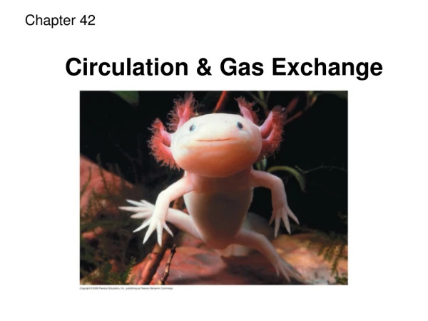
Chapter 42 Circulation and Gas Exchange
Open and Closed Circulatory Systems • Open circulatory system- (insects, arthropods, and mollusks) blood bathes the organs directly. Fluid is called hemolymph. • Closed circulatory system- blood is confined to vessels and is distinct from the interstitial fluid
Heart Heart Blood Hemolymph in sinuses surrounding organs Small branch vessels In each organ Interstitial fluid Pores Dorsal vessel (main heart) Tubular heart Auxiliary hearts Ventral vessels (a) An open circulatory system (b) A closed circulatory system
Organization of Vertebrate Circulatory Systems • Arteries branch into arterioles and carry blood to capillaries • Networks of capillaries (capillary beds) are the sites of chemical exchange between the blood and interstitial fluid • Venulesconverge into veins and return blood from capillaries to the heart • Blood enters through an atrium and is pumped out through a ventricle
Gill capillaries Single Circulation • Blood leaving the heart passes through two capillary beds before returning • Bony fishes, rays, and sharks have single circulationwith a two-chambered heart Gill circulation Artery Ventricle Heart Atrium Systemic circulation Vein Systemic capillaries
Double Circulation • Oxygen-poor and oxygen-rich blood are pumped separately from the right and left sides of the heart (Amphibian, reptiles, and mammals)
Adaptations of Double Circulatory Systems Amphibians- Frogs and other amphibians have a three-chambered heart: two atria and one ventricle Amphibians Reptiles (Except Birds) Mammals and Birds Lung and skin capillaries Lung capillaries Lung capillaries Right systemic aorta Pulmocutaneous circuit Pulmonary circuit Pulmonary circuit Atrium (A) Atrium (A) A A A A V V Ventricle (V) V V Left systemic aorta Left Right Left Right Right Left Systemic circuit Systemic circuit Systemic capillaries Systemic capillaries Systemic capillaries
Adaptations of Double Circulatory Systems • Reptiles (Except Birds)- Turtles, snakes, and lizards have a three-chambered heart: two atria and one ventricleIn alligators, caimans, and other crocodilians a septum divides the ventricle Amphibians Reptiles (Except Birds) Mammals and Birds Lung and skin capillaries Lung capillaries Lung capillaries Right systemic aorta Pulmocutaneous circuit Pulmonary circuit Pulmonary circuit Atrium (A) Atrium (A) A A A A V V Ventricle (V) V V Left systemic aorta Left Right Left Right Right Left Systemic circuit Systemic circuit Systemic capillaries Systemic capillaries Systemic capillaries
Adaptations of Double Circulatory Systems • Mammals and Birds • Mammals and birds have a four-chambered heart with two atria and two ventricles Amphibians Reptiles (Except Birds) Mammals and Birds Lung and skin capillaries Lung capillaries Lung capillaries Right systemic aorta Pulmocutaneous circuit Pulmonary circuit Pulmonary circuit Atrium (A) Atrium (A) A A A A V V Ventricle (V) V V Left systemic aorta Left Right Left Right Right Left Systemic circuit Systemic circuit Systemic capillaries Systemic capillaries Systemic capillaries
Capillaries of head and forelimbs Superior vena cava 7 Pulmonary artery Pulmonary artery Capillaries of right lung Aorta 9 Capillaries of left lung 3 3 2 4 11 Pulmonary vein Pulmonary vein 5 1 Right atrium Left atrium 10 Right ventricle Left ventricle Inferior vena cava Aorta Capillaries of abdominal organs and hind limbs 8 Animation: Path of Blood Flow in Mammals
The Mammalian Heart • Cardiac cycle- the rhythmic cycle of contracting and relaxing • Systole- the contraction, or pumping, phase • Diastole- the relaxation, or filling, phase • Four valves prevent backflow of blood in the heart • The atrioventricular (AV) valves separate each atrium and ventricle • The semilunar valves control blood flow to the aorta and the pulmonary artery
Pulmonary artery Aorta Pulmonary artery Right atrium Left atrium Semilunar valve Semilunar valve Atrioventricular valve Atrioventricular valve Right ventricle Left ventricle
Maintaining the Heart’s Rhythmic Beat • Sinoatrial(SA) node, or pacemaker, sets the rate and timing at which cardiac muscle cells contract • Impulses from the SA node travel to the atrioventricular (AV) node • At the AV node, the impulses are delayed and then travel to the Purkinje fibers that make the ventricles contract
3 1 2 Pacemaker generates wave of signals to contract. Signals are delayed at AV node. Signals pass to heart apex. Signals spread throughout ventricles. 4 SA node (pacemaker) AV node Purkinje fibers Bundle branches Heart apex ECG
Artery Vein SEM Valve 100 µm Basal lamina Endothelium Endothelium Smooth muscle Smooth muscle Connective tissue Connective tissue Capillary Artery Vein Arteriole Venule 15 µm Red blood cell Capillary LM
Blood Flow Velocity • Blood flow in capillaries is slow for the exchange of materials 5,000 4,000 Area (cm2) 3,000 2,000 1,000 0 50 40 Velocity (cm/sec) 30 20 10 0 120 Systolic pressure 100 80 Pressure (mm Hg) 60 Diastolic pressure 40 20 0 Aorta Veins Arteries Venules Arterioles Capillaries Venae cavae
Blood Pressure- Changes in Blood Pressure During the Cardiac Cycle • Systolic pressure is the pressure in the arteries during ventricular systole; it is the highest pressure in the arteries • Diastolic pressure is the pressure in the arteries during diastole; it is lower than systolic pressure
Regulation of Blood Pressure • Vasoconstriction is the contraction of smooth muscle in arteriole walls; it increases blood pressure • Vasodilationis the relaxation of smooth muscles in the arterioles; it causes blood pressure to fall
Blood Pressure and Gravity Direction of blood flow in vein (toward heart) • Blood is moved through veins by smooth muscle contraction, skeletal muscle contraction, and expansion of the vena cava with inhalation • One-way valves in veins prevent backflow of blood Valve (open) Skeletal muscle Valve (closed)
Capillary Function • The critical exchange of substances between the blood and interstitial fluid takes place across the thin endothelial walls of the capillaries Body tissue INTERSTITIAL FLUID Capillary Net fluid movement out Net fluid movement in Direction of blood flow
Fluid Return by the Lymphatic System • The lymphatic system returns fluid that leaks out in the capillary beds • Fluid, called lymph, reenters the circulation directly at the venous end of the capillary bed and indirectly through the lymphatic system • The lymphatic system drains into veins in the neck
Blood components function in exchange, transport, and defense • In invertebrates with open circulation, blood (hemolymph) is not different from interstitial fluid
Blood Composition and Function • Plasma- 90% water, inorganic salts (electrolytes), plasma proteins (influence blood pH, osmotic pressure, and viscosity also lipid transport, immunity, and blood clotting) • Erythrocytes- red blood cells, transport oxygen with hemoglobin • Leukocytes- white blood cells • Platelets- fragments of cells and function in blood clotting
Plasma 55% Constituent Major functions Cellular elements 45% Cell type Number per µL (mm3) of blood Functions Solvent for carrying other substances Water Erythrocytes (red blood cells) Transport oxygen and help transport carbon dioxide 5–6 million Ions (blood electrolytes) Sodium Potassium Calcium Magnesium Chloride Bicarbonate Osmotic balance, pH buffering, and regulation of membrane permeability Separated blood elements Leukocytes (white blood cells) Defense and immunity 5,000–10,000 Plasma proteins Albumin Osmotic balance pH buffering Lymphocyte Basophil Fibrinogen Clotting Eosinophil Immunoglobulins (antibodies) Defense Neutrophil Monocyte Substances transported by blood Nutrients (such as glucose, fatty acids, vitamins) Waste products of metabolism Respiratory gases (O2 and CO2) Hormones 250,000– 400,000 Platelets Blood clotting
Blood Clotting • A cascade of complex reactions converts fibrinogen to fibrin, forming a clot • A blood clot formed within a blood vessel is called a thrombus and can block blood flow Red blood cell Collagen fibers Platelet plug Fibrin clot Platelet releases chemicals that make nearby platelets sticky Clotting factors from: Platelets Damaged cells Plasma (factors include calcium, vitamin K) Prothrombin Thrombin Fibrinogen Fibrin 5 µm
Cardiovascular Disease • Arteriosclerosis- hardening of the arteries- can include atherosclerosis • Atherosclerosis- caused by the buildup of plaque deposits within arteries • Heart Attacks- the death of cardiac muscle tissue from blockage of coronary arteries • Stroke- the death of tissue in the brain, from rupture/blockage of arteries in the head
Smooth muscle Connective tissue Endothelium 50 µm (a) Normal artery
Plaque (b) Partly clogged artery 250 µm
Treatment and Diagnosis of Cardiovascular Disease • Low-density lipoproteins (LDLs) cause plaque formation; “bad cholesterol” • High-density lipoproteins (HDLs) reduce the deposition of cholesterol; “good cholesterol” • Hypertension, high blood pressure, increases the risk of heart attack and stroke
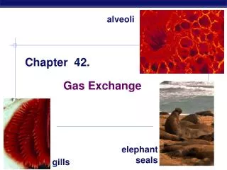
Gas exchange occurs across specialized respiratory surfaces • Gas exchange across respiratory surfaces takes place by diffusion Gills in Aquatic Animals • Fish gills use a countercurrent exchange system, where blood flows in the opposite direction to water passing over the gills; blood is always less saturated with O2 than the water it meets
Fluid flow through gill filament Oxygen-poor blood Anatomy of gills Oxygen-rich blood Gill arch Lamella Gill arch Gill filament organization Blood vessels Water flow Operculum Water flow between lamellae Blood flow through capillaries in lamella Countercurrent exchange PO2 (mm Hg) in water 150 120 90 60 30 Gill filaments Net diffu- sion of O2 from water to blood 110 80 50 20 140 PO2 (mm Hg) in blood
Tracheal Systems in Insects • The tracheal system of insects consists of tiny branching tubes that penetrate the body • The tracheal tubes supply O2 directly to body cells • The respiratory and circulatory systems are separate
Air sacs Tracheae External opening Tracheoles Mitochondria Muscle fiber Body cell Air sac Tracheole Trachea Body wall Air 2.5 µm
Mammalian Respiratory Systems • Air inhaled through the nostrils passes through the pharynx via the larynx, trachea, bronchi, bronchioles, and alveoli, where gas exchange occurs
Branch of pulmonary vein (oxygen-rich blood) Branch of pulmonary artery (oxygen-poor blood) Terminal bronchiole Nasal cavity Pharynx Larynx Alveoli (Esophagus) Left lung Trachea Right lung Bronchus Bronchiole Diaphragm Heart SEM Colorized SEM 50 µm 50 µm
Rib cage expands as rib muscles contract Rib cage gets smaller as rib muscles relax Air inhaled Air exhaled Lung Diaphragm INHALATION Diaphragm contracts (moves down) EXHALATION Diaphragm relaxes (moves up)
Control of Breathing in Humans Cerebrospinal fluid • The medulla oblongata regulates the rate and depth of breathing in response to pH changes in the cerebrospinal fluid. • The pons regulates the tempo • Sensors in the aorta and carotid arteries (secondary control) monitor O2 and CO2 concentrations in the blood Pons Breathing control centers Medulla oblongata Carotid arteries Aorta Diaphragm Rib muscles
Coordination of Circulation and Gas Exchange • In the alveoli, O2 diffuses into the blood and CO2 diffuses into the air • In tissue capillaries, partial pressure gradients favor diffusion of O2 into the interstitial fluids and CO2 into the blood
Respiratory Pigments • Arthropods and many molluscs have hemocyanin with copper as the oxygen-binding component • Most vertebrates and some invertebrates use hemoglobin • A single hemoglobin molecule can carry four molecules of O2 • CO2 produced during cellular respiration lowers blood pH and decreases the affinity of hemoglobin for O2; this is called the Bohr shift
Chains Iron Heme Chains Hemoglobin
Carbon Dioxide Transport • Hemoglobin also helps transport CO2 and assists in buffering • CO2 from respiring cells diffuses into the blood and is transported either in blood plasma, bound to hemoglobin, or as bicarbonate ions (HCO3–) Animation: O2 from Blood to Tissues Animation: CO2 from Tissues to Blood Animation: CO2 from Blood to Lungs Animation: O2 from Lungs to Blood