Overview of Circulatory Systems and Gas Exchange in Multicellular Organisms
This chapter explores the processes of circulation and gas exchange in organisms, highlighting the necessity of material exchange with the environment at the cellular level. For unicellular organisms, exchanges occur directly, while multicellular organisms rely on specialized systems, such as gills and circulatory systems. The chapter distinguishes between open and closed circulatory systems, detailing their components and efficiency in transporting blood. It also covers vertebrate cardiovascular systems, emphasizing double circulation in mammals and the heart's function in meeting oxygen demands.

Overview of Circulatory Systems and Gas Exchange in Multicellular Organisms
E N D
Presentation Transcript
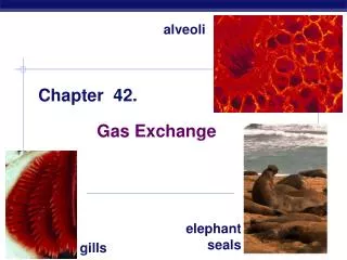
Chapter 42 Circulation and Gas Exchange
Overview: Trading Places • Every organism must exchange materials with its environment • Exchanges ultimately occur at the cellular level • In unicellular organisms, these exchanges occur directly with the environment
For most cells making up multicellular organisms, direct exchange with the environment is not possible • Gills are an example of a specialized exchange system in animals • Internal transport and gas exchange are functionally related in most animals
Concept 42.1: Circulatory systems link exchange surfaces with cells throughout the body • In small and/or thin animals, cells can exchange materials directly with the surrounding medium • In most animals, transport systems connect the organs of exchange with the body cells • Most complex animals have internal transport systems that circulate fluid
Gastrovascular Cavities • Simple animals, such as cnidarians, have a body wall that is only two cells thick and that encloses a gastrovascular cavity • This cavity functions in both digestion and distribution of substances throughout the body • Some cnidarians, such as jellies, have elaborate gastrovascular cavities • Flatworms have a gastrovascular cavity and a large surface area to volume ratio
Fig. 42-2 Circular canal Mouth Pharynx Mouth Radial canal 5 cm 2 mm (a) The moon jelly Aurelia, a cnidarian (b) The planarian Dugesia, a flatworm
Open and Closed Circulatory Systems • More complex animals have either open or closed circulatory systems • Both systems have three basic components: • A circulatory fluid (blood or hemolymph) • A set of tubes (blood vessels) • A muscular pump (the heart)
In insects, other arthropods, and most molluscs, blood bathes the organs directly in an open circulatory system • In an open circulatory system, there is no distinction between blood and interstitial fluid, and this general body fluid is more correctly called hemolymph
In a closed circulatory system, blood is confined to vessels and is distinct from the interstitial fluid • Closed systems are more efficient at transporting circulatory fluids to tissues and cells
Fig. 42-3 Heart Heart Blood Hemolymph in sinuses surrounding organs Small branch vessels In each organ Interstitial fluid Pores Dorsal vessel (main heart) Tubular heart Auxiliary hearts Ventral vessels (a) An open circulatory system (b) A closed circulatory system
Organization of Vertebrate Circulatory Systems • Humans and other vertebrates have a closed circulatory system, often called the cardiovascular system • The three main types of blood vessels are arteries, veins, and capillaries
Arteries branch into arterioles and carry blood to capillaries • Networks of capillaries called capillary beds are the sites of chemical exchange between the blood and interstitial fluid • Venules converge into veins and return blood from capillaries to the heart
Vertebrate hearts contain two or more chambers • Blood enters through an atrium and is pumped out through a ventricle
Single Circulation • Bony fishes, rays, and sharks have single circulation with a two-chambered heart • In single circulation, blood leaving the heart passes through two capillary beds before returning
Fig. 42-4 Gill capillaries Gill circulation Artery Ventricle Heart Atrium Systemic circulation Vein Systemic capillaries
Double Circulation • Amphibian, reptiles, and mammals have double circulation • Oxygen-poor and oxygen-rich blood are pumped separately from the right and left sides of the heart
Fig. 42-5 Amphibians Reptiles (Except Birds) Mammals and Birds Lung and skin capillaries Lung capillaries Lung capillaries Right systemic aorta Pulmocutaneous circuit Pulmonary circuit Pulmonary circuit Atrium (A) Atrium (A) A A A A V V Ventricle (V) V V Left systemic aorta Left Right Left Right Right Left Systemic circuit Systemic circuit Systemic capillaries Systemic capillaries Systemic capillaries
Concept 42.2: Coordinated cycles of heart contraction drive double circulation in mammals • The mammalian cardiovascular system meets the body’s continuous demand for O2
Mammalian Circulation • Blood begins its flow with the right ventricle pumping blood to the lungs • In the lungs, the blood loads O2 and unloads CO2 • Oxygen-rich blood from the lungs enters the heart at the left atrium and is pumped through the aorta to the body tissues by the left ventricle • The aorta provides blood to the heart through the coronary arteries
Blood returns to the heart through the superior vena cava (blood from head, neck, and forelimbs) and inferior vena cava (blood from trunk and hind limbs) • The superior vena cava and inferior vena cava flow into the right atrium
Fig. 42-6 Capillaries of head and forelimbs Superior vena cava 7 Pulmonary artery Pulmonary artery Capillaries of right lung Aorta 9 Capillaries of left lung 3 3 2 4 11 Pulmonary vein Pulmonary vein 5 1 Right atrium Left atrium 10 Right ventricle Left ventricle Inferior vena cava Aorta Capillaries of abdominal organs and hind limbs 8
Fig. 42-7 Pulmonary artery Aorta Pulmonary artery Right atrium Left atrium Semilunar valve Semilunar valve Atrioventricular valve Atrioventricular valve Right ventricle Left ventricle
Maintaining the Heart’s Rhythmic Beat • Some cardiac muscle cells are self-excitable, meaning they contract without any signal from the nervous system
The sinoatrial (SA) node, or pacemaker, sets the rate and timing at which cardiac muscle cells contract • Impulses from the SA node travel to the atrioventricular (AV) node • At the AV node, the impulses are delayed and then travel to the Purkinje fibers that make the ventricles contract
Impulses that travel during the cardiac cycle can be recorded as an electrocardiogram (ECG or EKG)
Fig. 42-9-5 3 1 2 Pacemaker generates wave of signals to contract. Signals are delayed at AV node. Signals pass to heart apex. Signals spread throughout ventricles. 4 SA node (pacemaker) AV node Purkinje fibers Bundle branches Heart apex ECG
Concept 42.3: Patterns of blood pressure and flow reflect the structure and arrangement of blood vessels • The physical principles that govern movement of water in plumbing systems also influence the functioning of animal circulatory systems
Fig. 42-10 Artery Vein SEM Valve 100 µm Basal lamina Endothelium Endothelium Smooth muscle Smooth muscle Connective tissue Connective tissue Capillary Artery Vein Arteriole Venule 15 µm Red blood cell Capillary LM
Capillaries have thin walls, the endothelium plus its basement membrane, to facilitate the exchange of materials • Arteries and veins have an endothelium, smooth muscle, and connective tissue • Arteries have thicker walls than veins to accommodate the high pressure of blood pumped from the heart • In the thinner-walled veins, blood flows back to the heart mainly as a result of muscle action
Blood Flow Velocity • Physical laws governing movement of fluids through pipes affect blood flow and blood pressure • Velocity of blood flow is slowest in the capillary beds, as a result of the high resistance and large total cross-sectional area • Blood flow in capillaries is necessarily slow for exchange of materials
Fig. 42-11 5,000 4,000 Area (cm2) 3,000 2,000 1,000 0 50 40 Velocity (cm/sec) 30 20 10 0 120 Systolic pressure 100 80 Pressure (mm Hg) 60 Diastolic pressure 40 20 0 Aorta Veins Arteries Venules Arterioles Capillaries Venae cavae
Blood Pressure • Blood pressure is the hydrostatic pressure that blood exerts against the wall of a vessel • In rigid vessels blood pressure is maintained; less rigid vessels deform and blood pressure is lost
Changes in Blood Pressure During the Cardiac Cycle • Systolic pressure is the pressure in the arteries during ventricular systole; it is the highest pressure in the arteries • Diastolic pressure is the pressure in the arteries during diastole; it is lower than systolic pressure • A pulse is the rhythmic bulging of artery walls with each heartbeat
Blood Pressure and Gravity • Blood pressure is generally measured for an artery in the arm at the same height as the heart • Blood pressure for a healthy 20 year old at rest is 120 mm Hg at systole and 70 mm Hg at diastole
Fig. 42-14 Direction of blood flow in vein (toward heart) Valve (open) Skeletal muscle Valve (closed)
Capillary Function • Capillaries in major organs are usually filled to capacity • Blood supply varies in many other sites
Two mechanisms regulate distribution of blood in capillary beds: • Contraction of the smooth muscle layer in the wall of an arteriole constricts the vessel • Precapillary sphincters control flow of blood between arterioles and venules
Fig. 42-15 Thoroughfare channel Precapillary sphincters Capillaries Arteriole Venule (a) Sphincters relaxed Arteriole Venule (b) Sphincters contracted
The critical exchange of substances between the blood and interstitial fluid takes place across the thin endothelial walls of the capillaries • The difference between blood pressure and osmotic pressure drives fluids out of capillaries at the arteriole end and into capillaries at the venule end
Fig. 42-16 Body tissue INTERSTITIAL FLUID Capillary Net fluid movement out Net fluid movement in Direction of blood flow Blood pressure Inward flow Pressure Outward flow Osmotic pressure Arterial end of capillary Venous end
Fluid Return by the Lymphatic System • The lymphatic system returns fluid that leaks out in the capillary beds • This system aids in body defense • Fluid, called lymph, reenters the circulation directly at the venous end of the capillary bed and indirectly through the lymphatic system • The lymphatic system drains into veins in the neck
Concept 42.4: Blood components function in exchange, transport, and defense • In invertebrates with open circulation, blood (hemolymph) is not different from interstitial fluid • Blood in the circulatory systems of vertebrates is a specialized connective tissue
Blood Composition and Function • Blood consists of several kinds of cells suspended in a liquid matrix called plasma • The cellular elements occupy about 45% of the volume of blood
Fig. 42-17 Plasma 55% Constituent Major functions Cellular elements 45% Cell type Number per µL (mm3) of blood Functions Solvent for carrying other substances Water Erythrocytes (red blood cells) Transport oxygen and help transport carbon dioxide 5–6 million Ions (blood electrolytes) Sodium Potassium Calcium Magnesium Chloride Bicarbonate Osmotic balance, pH buffering, and regulation of membrane permeability Separated blood elements Leukocytes (white blood cells) Defense and immunity 5,000–10,000 Plasma proteins Albumin Osmotic balance pH buffering Lymphocyte Basophil Fibrinogen Clotting Eosinophil Immunoglobulins (antibodies) Defense Neutrophil Monocyte Substances transported by blood Nutrients (such as glucose, fatty acids, vitamins) Waste products of metabolism Respiratory gases (O2 and CO2) Hormones 250,000– 400,000 Platelets Blood clotting
Plasma • Blood plasma is about 90% water • Among its solutes are inorganic salts in the form of dissolved ions, sometimes called electrolytes • Another important class of solutes is the plasma proteins, which influence blood pH, osmotic pressure, and viscosity • Various plasma proteins function in lipid transport, immunity, and blood clotting
Cellular Elements • Suspended in blood plasma are two types of cells: • Red blood cells (erythrocytes) transport oxygen • White blood cells (leukocytes) function in defense • Platelets, a third cellular element, are fragments of cells that are involved in clotting
Erythrocytes • Red blood cells, or erythrocytes, are by far the most numerous blood cells • They transport oxygen throughout the body • They contain hemoglobin, the iron-containing protein that transports oxygen
Leukocytes • There are five major types of white blood cells, or leukocytes: monocytes, neutrophils, basophils, eosinophils, and lymphocytes • They function in defense by phagocytizing bacteria and debris or by producing antibodies • They are found both in and outside of the circulatory system
Platelets • Platelets are fragments of cells and function in blood clotting