Download
1 / 23
230 likes | 452 Vues
Gene regulation overview and Saccharomyces cerevisiae cell-type specific mating type promoters. Nate Sotuyo Allison Suarez. Transcription. Prokaryotes. Overview. (RNA)n + ribonucleoside triphosphate ⇌ (RNA)n+1 +PPi Divalent metal ion required (Mg2+ orMn2+) Cytoplasm, mRNA isnot modified

E N D
Gene regulation overviewandSaccharomyces cerevisiae cell-type specific mating type promoters Nate Sotuyo Allison Suarez
Transcription Prokaryotes
Overview • (RNA)n + ribonucleoside triphosphate ⇌ (RNA)n+1 +PPi • Divalent metal ion required (Mg2+ orMn2+) • Cytoplasm, mRNA isnot modified • 5 Steps • Pre-initiation • Initiation • Promoter clearance • Elongation • termination
DNA Synthesis comparison Similarities • 5’ 3’ • Nucleophilic attack by 3’ OH on phosphate • Driven by hydrolysis or pyrophosphate • Pyrophosphate orthophosphate Differences • No Primer required • Error rate higher
Pre-initiation • RNA holoenzyme α2ββ'σω binds DNA • “slides” along double helix • Unwinds the DNA to form initiation bubble
Initiation and promoter clearance Initiation • RNA polymerase binds promoter (with sigma factor) • Prinow box (-10), -35 region • First phosphodiester bond formed Promoter clearance • Abortive initiation • CTD phosphorylation by TFIIH
Elongation and Termination Elongation • 5’3’, TU • σ factor released • TopoisomeraseII feedback Termination • Rho-independent • G-C hairpin followed by string of U’s • Rho-dependent • ρ destabilizes mRNA-template interaction
Review: Gene Regulation and Transcription in Eukaryotes • Similar to gene regulation in prokaryotes, except more complex: more types of regulatory proteins and interactions with regulatory regions in DNA • Both prokaryotes and eukaryotes have promoters • Region of DNA that is the RNA polymerase’s transcription initiation site • Important differences in eukaryotes: • Nucleosomes, chromatin • Chromatin remodeling • 3 types of RNA polymerases • RNA modification • Transcription occurs in the nucleus • Translation occurs in the cytoplasm • Binding of DNA-binding proteins outside the promoter region
Example of chromatin remodeling • Chromatin remodeling is the changing of nucleosome position • Histones • Core octamer, amino-terminal ends • Covalent modification termed “histone code” • Acetylation, deacetylation, and gene expression • Acetylation of certain histone residues can cause the histone to slide along the DNA, no longer inhibiting a promoter region or other important DNA-protein binding sites necessary for transcription
Overview of transcription translation in eukaryotes http://www1.ci.uc.pt/yss/Material/pdf_RNA%20Regulation/figurea6.jpg
Genomic Footprinting of the Promoter Regions of STE2 and STE3 genes in the Yeast Saccharomyces cerevisiae Brigitte Ganter, Song Tan and Timothy J. Richmond
Overview Overall goal • Develop high resolution map of the promoter regions of STE2 and STE3 • Analyze the chromatin structure in the promoter regions using DMS methylation, micrococcal nuclease and DNase I.
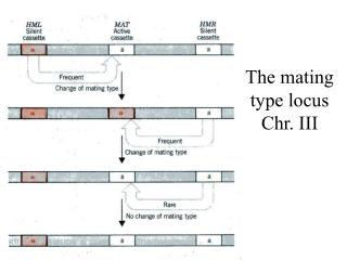
Mating types review • Three cell types • Haploid: a,α • Diploid: a/α • Specific gene expression determined by MATa or MATα • Three transcription regulators expressed • MATa1,MATα1,MATα2 • Other key transcription factors • MCM1, STE12, SSN6, TUP1
Transcription factors • MCM1 • Activates both a and alpha • Purified vs. crude extracts (Q factors?) • a-specific genes conformational change • STE12 • Component of pheromone response pathway • Binds PRE signaling cascade • Poor interaction unless MCM1 present • MATα2 • Precise positioning of nucleosomes downstream STE2 UAS elements in α cells
Transcription factors cont. • SSN6 and TUP1 • Required for full repression of a-genes in alpha cells • Mutated ssn6 resulted in alpha cells that could mate with other alpha cells (Though not efficiently)
Haploid Mating overview STE3 STE2 http://www.ncbi.nlm.nih.gov/books/bv.fcgi?highlight=p-box&rid=mcb.section.3752#3756
http://www.ncbi.nlm.nih.gov/books/bv.fcgi?highlight=p-box&rid=mcb.section.3752#3756http://www.ncbi.nlm.nih.gov/books/bv.fcgi?highlight=p-box&rid=mcb.section.3752#3756
Diploid Mating overview http://www.ncbi.nlm.nih.gov/books/bv.fcgi?highlight=p-box&rid=mcb.section.3752#3756
Significant nucleosome positioning • Contribute to transcription regulation • Stably positioned nucleosomes STE2/ α cells • Only MCM1 bound no stable array • TFIID binding site is protected • a-cells only protected in TATA region • Due to TFII binding, not nucleosome
Nucleosomal protection cont. • STE 3 more complicated • a-cells • MCM1 binding to P’-box on coding strand only in vitro • in vivo result consistent with each other • alpha-cells • Concerted binding of MCM1 and MAT-alpha1 • Additional protection not detected in vitro observed on Q-box side • Possible other factors that bind to these regions
Ligation Mediated PCR “Conventional PCR is not immediately applicable to sequencing or footprinting because it requires two defined ends. A sequence or footprint ladder is composed of a population of related nucleic acid fragments. One end of each fragment is fixed by a primer or restriction cut and is therefore the same for all, whereas the other end is determined by variable chemical cleavage or chain termination and is therefore unique for each fragment. To apply PCR to a sequence ladder, [Mueller and Wold] introduced a simple ligation step that adds a common oligonucleotide sequence to the unique end of each member. A primer complementary to this new common sequence is then used, together with a primer complementary to the original fixed end, for simultaneous exponential amplification of all members of the sequence ladder. The procedure has high selectivity and specificity that are derived from the design of the ligation step and choice of primers. It also has high fidelity; a footprint consists of subtle differences in the starting concentrations of particular members of a sequence a sequence ladder, and these differences are reproducibly retained through amplification.” Mueller, P.R. and Wold, B. (1989). In vivo footprinting of a muscle specific enhancer by ligation mediated PCR. Science, 246, 280-286
Mueller, P.R. and Wold, B. (1989). In vivo footprinting of a muscle specific enhancer by ligation mediated PCR. Science, 246, 280-286