Download
1 / 29
290 likes | 458 Vues
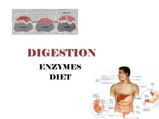
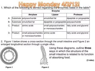
Happy Monday 4/2/12!. 1. Which of the following is correct regarding the enzymes listed in the table?. 2. Figure 1 below shows a cross-section through the small intestine and Figure 2 an enlarged longitudinal section through a single villus.

E N D
Happy Monday 4/2/12! 1. Which of the following is correct regarding the enzymes listed in the table? 2. Figure 1 below shows a cross-section through the small intestine and Figure 2 an enlarged longitudinal section through a single villus. Using these diagrams, outline three ways in which the structure of the small intestine is related to its function of absorbing food. (3 mks) Figure 1Figure 2
A • villus intestinal wall has many folds to increase surface area ( : volume ratio); surface of villus close to blood vessels so materials can easily diffuse; surface of villus close to lymph vessels so lipids can be easily absorbed; greater surface area related to greater rate of diffusion;villus wall consists of single layer of cells; • 3 maxDo not accept microvilli – not visible in diagrams. • The intestinal wall of the villus has many folds to increase surface area, or the SA:Vol. This increased surface area is related to an increased rate of diffusion. Aiding in diffusion is the fact that the wall of the villus consists only of a single layer of cells. The surface of the villus itself is located close to the blood vessels, so that materials can easily diffuse into them. It is also located close to the lymph vessels, so that lipids can easily be absorbed.
6.1.1 Explain why digestion of large food molecules is essential. • Most food molecules are large polymers & insoluble • Too big to pass through membrane or into blood • Must be digested smaller , soluble molecsabsorbed to blood • Series of chem reactions smaller & smaller molecules • Protein amino acids • Example: you eat egg albumin (egg white) contains amino acid serine hydrolysis reactions break down prtn into aa’s serine absorbed @ small intestine capillary, bloodstream pancreas cell Transcription/Translation used to make a new protein hormone insulin molecule • Lipids (triglycerides) glycerol, fatty acids • Carbs (poly/di/monosaccharides) monosaccharides • Nucleic acids (DNA/RNA) nucleotides • THEN, can be absorbed through cells of dig. system and blood • Circulatory sys transports nutrients to body cells • Body uses to make your “own” DNA, protein, etc.
6.1.2 Explain the need for enzymes in digestion. • Enzymes = biol catalysts, increase rate of reaction • Increase rate of digestion at body temperature! • Activation energy reduced: normal rxn – higher act energy –higher body temp----not possible in humans! • Digestive enzymes specific for specific food types • Secreted into lumen of gut • Hydrolysis of insoluble food molec into soluble products • Break down macromolecules • Starts @ mouth—salivary amylase • Starch (amylose) maltose glucose
6.1.3 State the source, substrate, products and optimum pH conditions for one amylase, one protease and one lipase. • Any human enzymes can be selected. Details of structure or mechanisms of action are not required. • Pancreatic amylase: • Source: pancreas cells • Optimal pH 7.5 – 7.8 • Substrate: starch (amylose) • End product: maltose (disaccharide) • Action: hydrolysis of 1-4 glycosidic bonds
6.1.3 State the source, substrate, products and optimum pH conditions for one amylase, one protease and one lipase. • Pepsin: (a protease) • Source: stomach cells • Optimal pH 2 - 3 • Substrate: polypeptide chains of amino acids • End product: amino acids • Action: hydrolysis of peptide bonds
6.1.3 State the source, substrate, products and optimum pH conditions for one amylase, one protease and one lipase. • Pancreatic lipase: • Source: pancreas cells • Optimal pH 7 • Substrate: triglyceride lipid • End product: glycerol & fatty acid chains • Action: requires bile salts to emulsify lipids (increases SA of lipid, for fat digestion, exposes glycerol structure to enzyme • Hydrolysis of ester bonds b/w glycerol and fatty acids
6.1.4 Draw and label a diagram of the digestive system. • “Alimentary Canal” • mouth, esophagus, stomach, small intestine, large intestine, anus, liver, pancreas and gall bladder. • The diagram should clearly show the interconnections between these structures!!!!!
6.1.5 Outline the function of the stomach, small intestine and large intestine. 1. Stomach: @ end of esophagus (peristalsis, muscular) Stores food, begins protein digestion. (a) Lumen: mixes food w/gastric juice (pepsin, HCl, mucus) (b) Gastric pits: mucus , enzymes and acid are secreted (c) Mucus secreting cells. (protect surface of stomach from auto-digestion) (d) Parietal cells: produce HCl which kills microorganisms that enter the digestive system (food & tracheal mucus). This also converts inactive pepsinogen to active pepsin & helps degrade foods some. (e) Chief cells: produces pepsinogen (protease enzyme) activated by HCl pepsin, most active in acidic pH
6.1.5 Outline the function of the stomach, small intestine and large intestine. 2. small Intestine: wheredigestion is completed & products of digestion are absorbed into the blood stream Duodenum: bile from liver/gall bladder trypsin (protease), lipase, amylase, bicarbonatefrom pancreas • Small molecules absorbed 1000s of villi (each w/cap bed and lacteal) increase Sfc Area Most molecules absorbed @ cap bed Lipids absorbed @ lacteal To body cells via another capillary & used for energy or building new macromolecules (assimilation) (a) Villus: increase SA for absorption of products of digestion (b) Microvilli: border of epithelial cell, increase the SA for absorption. (c) Lacteals connect to lymphatic system for the transport of lipids (d) In the wall of the small intestine are the blood vessels to transport absorbed products to the general circulation, And muscles to maintain peristalsis
6.1.5 Outline the function of the stomach, small intestine and large intestine. 3. Large Intestine or colon: responsible for reabsorption of water from gut. Undigested food (unabsorbed) excreted Water we drink/eat – much is reabsorbed, some passes E. coli (mutualistic) synthesize Vitamin K for us! (a) The lumen of the colon (b) The mucus producing goblet cells (b) Muscular walls to maintain peristalsis
6.1.6 Distinguish between absorption and assimilation. Insoluble food molecules are digested to soluble products in the lumen of the gut. Absorption: • soluble products are 1st taken up by various mechanisms into epithelial cells lining gut. • These cells then load various absorbed molecules into blood stream. Assimilation: • soluble products of digestion then transported to various tissues by circulatory system. • cells of tissues then absorb molecules for use within tissues
6.1.7 Explain how the structure of the villus is related to its role in absorption and transport of the products of digestion. • structure increases SA for absorption of digested food molecules into circ/lymph systems for transport • Digested nutrients from lumen of small int pass through single-cell thickness of villus epithelium to get to cap bed or lacteal • Undigested nutrients can’t pass through • Blood/lymph from arteriole/lacteal enters villus, picks up nutrients, leaves via venule/lacteal (a) folds increase SA:VOL ratio by X 3 (b) Villi project into the lumen of the gut increasing the surface area by X 10 (c) Microvilli are outward folds of the plasma membrane increasing the surface area another X10
6.1.7 Explain how the structure of the villus is related to its role in absorption and transport of the products of digestion. • Blood supply in villusabsorbs end products of digestion from epithelial cells • lacteals receive lipoproteins before transporting to circulatory system.