Download

1 / 25
250 likes | 377 Vues
This project aims to create a collaborative problem-solving environment in medical informatics to support healthcare professionals in diagnosing and managing breast cancer. It focuses on the development of user-friendly image registration services to establish spatial correspondence between contrast-enhanced breast MRI images. The integrated service incorporates validated workflows and accessibility across hospitals, utilizing advanced grid computing for efficient processing. The ultimate goal is to improve detection, diagnosis, and management of breast cancer while ensuring security and fault tolerance in communications.

E N D
Designing Grid-Enabled Image Registration Services for MIAKT Yalin Zheng, Christine Tanner, Derek L. G. Hill, David J. Hawkes Division of Imaging Sciences, King's College London, UK
MIAKT • Aim: to develop a collaborative problem solving environment in medical informatics • Application: support surgeons, radiologists, pathologists and oncologists in the detection, diagnosis and management of breast cancer
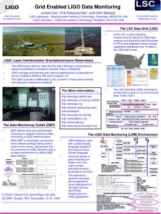
Image Registration • To establish spatial correspondence between images and possibly physical space • Application: Contrast-enhanced breast MRI pre-contrast post-contrast difference after registration
Image registration of contrast-enhanced breast MR images • User friendly • Integrated with other standard assessment tools • Accessible from many hospitals • Acceptable response time for a small number of cases • Consistent and well validated
Image Registration Technique • Non-rigid registration is based on evenly spaced control points interpolated by B-Splines Rueckert D. et al, IEEE TMI, vol. 18(8), pp. 712-721, 1999. • Accuracy for registering contrast-enhanced breast MR images has been carefully validated Schnabel J. et al, IEEE TMI, vol. 22(2), pp. 238-247, 2003. Tanner C. et al, MICCAI02, pp. 307-314, 2002. http://www-ipg.umds.ac.uk http://www.imageregistration.com
Workflow • A simple and validated workflow is provided for registering breast MR images • Individual registration of left and right side • Lesion alignment: rigid registration followed by non-rigid registration with 40mm control point spacing (~0.5 hours) • Whole breast alignment: 10mm multi-resolution registration (rigid -> 20mm non-rigid -> 10mm non-rigid) (~2.8 hours) • Optimal parameter as defaults • Makes service consistent and user-friendly
Workflow as above
Response Time Requirement • 30 minutes to 3 hours per side x 2 sides x 5 images = 5 hours to 30 hours on single machine • BUT, want results within hours not days • 10 individual jobs -> distribute them on 10 machines • Condor 6.4.5 • Distributes jobs to available machines • Job priority and dependency • Fault tolerance (checkpointing, rollback recovery)
Accessibility Requirement • Many hospitals • Service and images at different sites • Globus Toolkit 2.4 • Security • Resource Management • Combine condor and globus (Condor-G)
Security Requirement • Communications among organizations over Internet are essential for Grid applications, but Internet may be untrusted • Globus with Firewall • Configurations on firewall are required on both Client and Server sides to allow communications between them • Issues: • System admin should gain experiences for open ports required by Globus, e.g., 2811, 2119 and others • Great care should be taken to ensure security - Von Welch ,Globus Toolkit Firewall Requirements
Integrated Service • Tomcat Web Server, JSP/Servlet • WSDL file defines registration service • Clients can dynamically invoke the services • Registration submission • Registration monitoring • MIAKT calls it via SOAP through a Web-Service invocation architecture
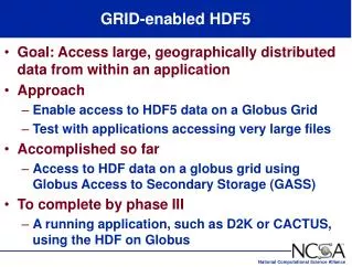
Configuration Overview Condor Submit Machine Globus Server (Job Manager, GSI-FTP etc.) Tomcat Web Server Engine
Successfully integrated with MIAKT demonstrator (Southampton booth)
Multi-Centre Trial • MARIBS: to test if MRI is an effective way of screening young women with high risk of breast cancer • 1500 women (35-49 yrs old) with high breast cancer risk • Annual MRI as well as X-ray mammograms for up to five years • 17 major screening centres
Segmentation refinement and classification of MR breast lesions
suspicious Intensity benign malignant Time Classification of MR Breast Lesions pre post-pre Features • Shape • Margins • Enhancement Pattern • Contrast-change Characteristics Time
Motivation • Segmentation Refinement • Feature extraction requires segmentation of MR breast lesion • Manual segmentation labour-intensive and difficult for 4D data • Derive most probable region from crude outline of lesion • Overall • Support radiologists in the diagnosis of MR breast lesions • Ease creation of large databases of annotated MR breast lesions with known ground truth (pathology / follow up)
Functionality & Design • Extract most probable region from 4D data of crude outline • Tanner C., MICCAI, September 2004 • Derive features from segmented region • Classifier: Linear discriminate analysis and leave-one-out ROC training • Online retraining of classifier • MATLAB program called from a Tomcat Web-Service implementation
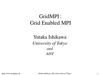
Results • Segmentation • Classification Accuracy • 10 benign, 16 malignant cases from MARIBS data set • 69% for features from gold standard segmentation • 82% for features from refined segmentation Initial Refined Gold Standard pre post1-pre post4-pre
Result MIAKT demonstrator
Conclusions • KCL services • GRID-enabled Image Registration Service • Segmentation Refinement and Classification Service for MR Breast Lesions • Services were successfully integrated with the MIAKT demonstrator • Future: from demonstrator to clinical usability
Thank you! • Funding from EPSRC • MIAKT • MIAS-IRC • MIAKT collaborators • University of Southampton • Sri Dasmahapatra, David Dupplaw, Bo Hu, Hugh Lewis, Paul Lewis, Nigel Shadbold, • University of Sheffield • Kalina Bontcheva, Fabio Ciravegna, Yorick Wilks • Open University • Liliana Cabral, John Domingue, Enrico Motto • University of Oxford • Mike Brady, Maud Poissonnier • Clinical Support • Guy’s Hospital London • Nick Beechy-Newman, Corrado D’Arrigio, Annette Jones