Download

1 / 42
500 likes | 711 Vues
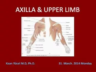
Axilla. Dr. K. S. Ravi MBBS, MD(JIPMER), MAMS Department of Anatomy AIIMS Rishikesh. Objectives. Definition of Axilla Boundaries Cervico-Axillary Canal Spaces/ Gateways in posterior wall Axillary folds & Sheath Contents of Axilla Axillary artery- Parts & Branches

E N D
Axilla Dr. K. S. Ravi MBBS, MD(JIPMER), MAMS Department of Anatomy AIIMS Rishikesh
Objectives • Definition of Axilla • Boundaries • Cervico-Axillary Canal • Spaces/ Gateways in posterior wall • Axillary folds & Sheath • Contents of Axilla • Axillary artery- Parts & Branches • Axillary Vein & Lymph nodes • Applied Anatomy
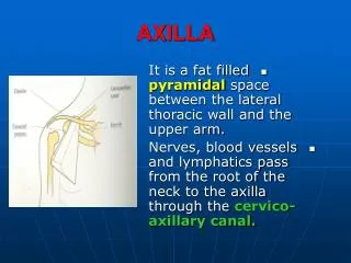
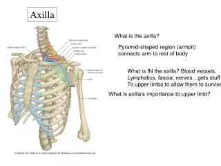
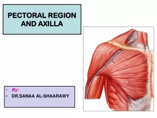
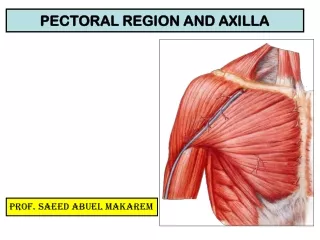
Axilla (arm pit) • Axilla is gateway to upper limb, providing an area of transition between neck & arm • Between upper arm & thoracic wall • Boundaries • -Apex • -Base • -Anterior wall • -Posterior wall • -Medial wall • -Lateral wall
What is Axilla? • A region (the axillary space) associated with armpit. • It actually begins around cervicoaxillary canal, at edge of first rib. • It continues to armpit, with bottom being axillary fascia. (remember? The lower attachment of the clavipectoral membrane?) • It has musculoskeletal boundaries that are lateral, medial, anterior and posterior.
MUSCLES Major structures inside: Axillary sheath and contents! Subscapularis M Latissimus dorsi M Teres major M Most of the rest of the space is adipose tissue. Pectoralis major M Serratus anterior M Pectoralis minor M
Apex/cervico-axial canal/Inlet • Directed towards the root of the neck • Boundaries: • Front: clavicle • Behind: upper border of scapula • Medially: outer margin of 1st rib • Transmits axillary vessels and brachial • plexus
Anterior wall: • pectoralis major • pectoralis minor • subclavius • clavipectoral fascia
Posterior wall: • Scapula • Subscapularis • Teres major • Latissmusdorsi (forms posterior axillary fold
Gateways in the posterior wall • Quadrangular space • provides a passageway for nerves and vessels passing between the axilla and the more posterior scapular and deltoid regions • Boundaries are formed by: • the inferior margin of the subscapularis/teres minor muscles • the surgical neck of the humerus; • the superior margin of the teres major muscle • axillary nerve and posterior circumflex humeral artery and vein passes through it
Gateways in the posterior wall • Triangular spaces • Upper triangular space: When viewed from anteriorly, it is formed • by: • Medial margin of the long head of the triceps brachii • Superior margin of the teres major muscle; • Inferior margin of the subscapularis/teres minor muscles. • Circumflex scapular artery & veinpass into this space.
Gateways in the posterior wall • Lower triangular interval is formed by: • long head of the triceps brachii muscle; • the shaft of the humerus; • the inferior margin of the teres major muscle • The radial nerve passes out of the axilla traveling through this interval to reach the posterior compartment of the arm.
Medial wall: • upper 3 or 4 intercostal spaces • upper part of serratus anterior • long thoracic nerve • intercosto brachial nerve pierces the medial wall
Lateral wall: • bicipital groove/intertubercular sulcus of the humerus • coraco brachialis • biceps brachii
Base: -skin -superficial fascia -axillary fascia (deep fascia)
Axillary folds: • Anterior axillary fold: • Pectoralis major • Posterior axillary fold: • Latissimusdorsi • Teres major
Contents: -axillary vessels -cords and branches of brachial plexus -axillary lymph nodes -fat
Axillary sheath • Derived, at least in part, from anterior and middle scalene muscle fascia. • Covers over a series of contents: • Axillary artery • Axillary vein • Brachial plexus and nerves derived from it. • The axillary sheath is just the fascia surrounding these structures.
Vertebral Artery Branches you should know: Transverse cervical. Dorsal scapular. Suprascapular. Subclavcian Artery. Lateral to the first rib, it becomes axillary artery.
Axillary Artery: divided into three parts Subclavian A. Part 1 (proximal) one branch Part 2 (intermediate) two branches. Brachial A. Part 3 (distal) three branches.
Axillary Artery: First Part From lateral border of 1st rib to medial border of Pectoralis Major M. Named Branch: Supreme Thoracic A. (to external thoracic body wall) Supplies blood to first and second intercostal spaces
Axillary Artery: Second part Deep to the pectoralis minor M. Thoracoacromial trunk Branches to: Clavicular area Pectoralis region Acromion of Scapula Deltoid Muscle. Lateral Thoracic Artery Bbr. to Serratus Ant. M.
Axillary Artery: third part Lateral border of Pectoralis minor M. to lateral border of Teres major M. Subscapular A.: Branches: Posterior circumflex humeral A. Circumflex scapular A. (to multiple muscles associated with the scapula) 1. Anterior circumflex humeral A. Thoracodorsal A. (to Latissimus dorsi M.) 2. How it will look in lab
Thoracoacromial A. Lateral thoracic A. Supreme thoracic A. Subscapular A. Post. Circumflex humoral A. Ant. Circumflex humoral A. Note, there is a broad anastamosis of the entire scapular region including circumflex humorals, subscapular, dorsal scapular, and suprascapular AA.
Arteries of Proximal Arm • The arterial pattern has one major vessel, with several important branches, which can supply muscles: • Deep brachial A. to posterior compartment (branches to medial collateral and radial collateral AA). • Superior ulnar collateral A. • Inferior ulnar collateral A. • Note, many muscles are supplied directly by unnamed muscular branches. Do not even think of giving all the vessels you see a distinct name.
The brachial artery is the primary artery supplying muscles of the arm. Note, the muscles and overlying skin are supplied by small, otherwise unnamed branches arising from it. Its largest single branch, the deep brachial A., arises from it in the upper part of the arm and penetrates towards the extensor (posterior) compartment. There are also arteries that supply the elbow anastomosis arising from it.
Axillary A. Brachial A. Deep brachial A. Radial collateral A. (a branch of the deep brachial A.) Superior ulnar collateral A. Not seen, middle collateral A., another branch of the deep brachial A. Inferior ulnar collateral A.
Axillary vein: Formation: Continuation of basilic vein at the lower border of teres major - runs upwards on the medial side of the axillary artery -Ends at outer border of 1st rib by becoming the subclavian vein Tributaries: -cephalic vein -veins corresponding to the branches of axillary artery
Vena comitans • Frequently multiple(2 or3) • Run with their corresponding • Arteries
Axillary sheath: axillary vessels and brachial plexus enclosed by a sheath of fascia called axillary fascia -continuous with the prevertebral fascia at the root of the neck
Axillary lymph nodes • There about 20 to 30 axillary lymph modes • Arranged in 5 groups: • 1.Anterior(pectoral) • 2.Posterior(subscapular) • 3.Lateral • 4.Central • 5.Apical
Regional Lymph Nodes for Breast A: Pectoralis major muscle B: Axillary lymph nodes level I C: Axillary lymph nodes level II D: Axillary lymph nodes level III E: Supraclavicular lymph nodes F: Internal mammary lymph nodes
?? 1. What are the structures that make up anterior wall of the axilla? • Pectoralis major, clavipectoral fascia and pectoralis minor • Subscapularis, teres major and latissimusdorsi • Pectoralis major, clavipectoral fascia and latissimusdorsi • Subscapularis, teres major and pectoralis minor
2. What are the structures that make up the posterior wall of the axilla? • Pectoralis major, clavipectoral fascia and pectoralis minor • Subscapularis, teres major and latissimusdorsi • Pectoralis major, clavipectoral fascia and latissimusdorsi • Subscapularis, teres major and pectoralis minor
3. The anterior group of axillary lymph nodes lies along the: • Lower border of the pectoralis minor • Lower margin of the posterior wall • Posteromedial to the axillary vein • In the fat of axilla • Behind and above the pectoralis minor, medial to the axillary vein
4. The apical group of axillary lymph nodes lies along the: • Lower border of the pectoralis minor • Lower margin of the posterior wall • Posteromedial to the axillary vein • In the fat of axilla • Behind and above the pectoralis minor, medial to the axillary vein
5. Which of the following branches of the axillary artery supplies the pectoral muscles and the thoracic wall? • Anterior circumflex humeral artery • Lateral thoracic artery • Posterior circumflex humeral artery • Superior thoracic artery
6. Which of the following branches of the axillary artery supplies the head of the humerus and the shoulder joint? • Anterior circumflex humeral artery • Lateral thoracic artery • Posterior circumflex humeral artery • Superior thoracic artery
Thank You