Download
1 / 56
1.01k likes | 2.48k Vues
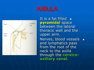
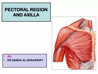
DISSECTION OF THE PECTORAL REGION AND AXILLA Make certain you have reviewed the anatomy of the brachial plexus the branches of the axillary artery the anatomy of the superficial veins in this region!!!. B. C. A. B. BRACHIAL PLEXUS. Anterior. Anterior. Post. Anterior. Post.

E N D
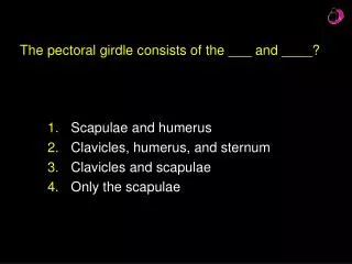
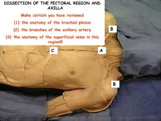
DISSECTION OF THE PECTORAL REGION AND AXILLA • Make certain you have reviewed • the anatomy of the brachial plexus • the branches of the axillary artery • the anatomy of the superficial veins in this region!!! B C A B
BRACHIAL PLEXUS Anterior Anterior Post. Anterior Post. Posterior Nerves Cords Divisions Trunks Rami Musculocut. C5 Lateral Upper C6 Median Middle C7 Medial C8 Ulnar Lower T1 Radial Posterior Axillary
ANT. ANT. POST. POST. POST. ANT. Brachial Plexus C5 Trunks C6 C7 SUPERIOR Divisions C8 Cords MIDDLE T1 INFERIOR Nerves LATERAL POSTERIOR Rami MUSCULOCUT. AXILLARY MEDIAL RADIAL MEDIAN . ULNAR
Superior thoracic Thoraco- acromial Lateral thoracic Axillary Anterior circumflex humeral Subclavian Posterior circumflex humeral Subscapular Catheter Circumflex scapular Thoracodorsal Deep brachial Axillary Artery Brachial
Veins Cephalic Axillary Brachial Basilic
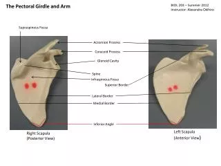
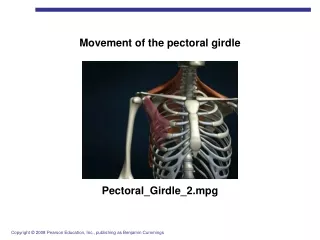
PECTORAL SKIN INCISIONS • Using the following slide as a reference, make the following skin incisions with the cadaver in the supine position. • From the jugular notch A along the clavicle and across the acromion B to a point about 10 cm distal to the acromion. • From A to the xiphisternal junction C. • From C laterally to the table. • At about mid-arm, make a complete circular incision. • At the level of the wrist make another circular incision. • Join these two circular incisions with a longitudinal one on the lateral aspect of the upper limb, that extends to the cut that is distal to the acromion.. Reflect the skin of the arm and forearm and remove it completely. DO NOT damage the superficial veins and cutaneous nerves in the superficial fascia.
PECTORAL REGION B C A B
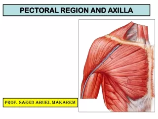
Clean the anterior surface of the pectoralis major muscle and define its borders. Recall that the pectoralis major muscle has two heads: a clavicular and sternocostal head. The fibers of the two heads converge and insert into the lateral lip of the bicipital groove of the humerus.
Identify the cephalic vein piercing the fascia of the deltopectoral triangle.
When beginning dissection in this region, recognize and SAVE the thoracoacormial trunk or artery as it exits the axillary artery
Cut the sternocostal head of pectoralis major muscle near its attachment to the sternum and the costal cartilage of the 6th rib and reflect it laterally.
Lateral pectoral nerve Pectoralis minor m. Medial pectoral nerve The medial and lateral pectoral nerves are named for the cords of the brachial plexus from which they arise and not for their relationship topectoralis minor m.. You should note some branches of the thracoacromial artery accompanying these nerves. Serratus anterior m.
ANT. ANT. POST. POST. POST. ANT. Brachial Plexus C5 C6 C7 SUPERIOR Lateral pectoral nerve C8 MIDDLE T1 INFERIOR LATERAL POSTERIOR MUSCULOCUT. AXILLARY MEDIAL RADIAL Medial pectoral nerve MEDIAN . ULNAR
Identify the borders of the pectoralis minor m. Cut it near its attachments to the ribs and reflect the muscle superiorly towards its attachment to the coracoid process.
As you reflect the pectoralis minor m., you should be able to better view the medial and lateral poctoral nn. And the accompanying vessels, which are branches of the thoracoacromial artery. Remember that the thoracoacromial a. arises from the second part of the axillary a., which is the portion of the vessel that lies deep to the pectoralis minor m. Thorcoacromial a.
This is a good time to also identify and save the lateral thoracic a., which also comes off the second part of the axillary a.. It can be found along the lateral border of the pectoralis minor m. accompanied by the long thoracic n. which innervates the serratus anterior m.. Subclavius m. Lateral thoracic a. Long thoracic n.
ANT. ANT. POST. POST. POST. ANT. Brachial Plexus Long thoracic nerve C5 C6 C7 SUPERIOR C8 MIDDLE T1 INFERIOR LATERAL POSTERIOR MUSCULOCUT. AXILLARY MEDIAL RADIAL MEDIAN . ULNAR
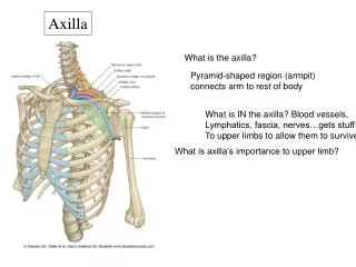
The axilla is described anatomically as having four walls. Anterior – clavicle and pectoral mm. Lateral – humerus. Medial – ribs and intercostal mm.. Posterior – scapula and subscapularis m.
The axilla is described anatomically as having four walls. Anterior – clavicle and pectoral mm. Lateral – humerus. Medial – ribs and intercostal mm.. Posterior – scapula and subscapularis m.
Make certain that the arm is in an abducted position to facilitate the cleaning of the nerves and vessels. Be careful to NOT cut the nerves and blood vessels contained in the axilla as the superficial fascia is removed.
Now, clean and identify the terminal branches of the brachial plexus.
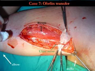
Musculocutaneous nerve – arising from the lateral cord and piercing the coracobrachialis muscle Coracobrachialis muscle
Contribution from lateral cord Contribution from medial cord Median nerve Recall that the median nerve has contributions from both the lateral and medial cords of the brachial plexus.
You will notice two nerves running adjacent to the ulnar nerve that may arise separately from the medial cord or from a common trunk – the medial brachial cutaneous and medial antebrachial cutaneous nerves. These nerves provide cutaneous innervation to the medial aspect of the arm and forearm. Medial cutaneous nerve of the arm or medial brachial cutaneous nerve Medial cutaneous nerve of the forearm or medial antebrachial cutaneous nerve
ANT. ANT. POST. POST. POST. ANT. Brachial Plexus C5 C6 C7 SUPERIOR C8 MIDDLE T1 INFERIOR LATERAL POSTERIOR MUSCULOCUT. AXILLARY MEDIAL RADIAL Medial brachial cutaneous nerve MEDIAN . ULNAR Medial antebrachial cutaneous nerve
You may also see the intercostobrachial nerve which actually arises from the second or third intercostal nerves.It will often communicate with the medial brachial cutaneous nerve. The intercostobrachial nerve primarily supplies the skin on the floor of the axilla and some of the adjacent regions of the arm. Intercostalbrachial nerve
Lateral pectoral nerve arising from the lateral cord Medial pectoral nerve arising from the medial cord You should be able to trace the medial and lateral pectoral nerves to their origins from the medial and lateral cords of the brachial plexus.
Now, let’s take another look at the axillary arterywhichbegins at the lateral border of the first rib as the continuation of the subclavian artery. It is described as being made up of three parts relative to the pectoralis minor muscle.
The first part of the axillary artery lies between the lateral border of the first rib and the medial border of pectoralis minor muscle.
reflected pectoralis minor muscle The second part of the axillary artery lies posterior to the pectoralis minor muscle.
The third part of the axillary artery lies between the lateral border of the pectoralis minor muscle and inferior border of teres major muscle. As the axillary artery passes distal to the teres major muscle, its name changes to the brachial artery.
BRANCHES OF THE AXILLARY ARTERY First Part (located between lateral border of first rib and superior border of pectoralis minor) 1. superior thoracic artery Second Part (deep to the pectoralis minor muscle) 1. thoracoacromial trunk or artery 2. lateral thoracic artery Third Part ( from inferior border of the pectoralis minor muscle to the inferior border of the teres major muscle) 1. subscapular artery 2. anterior circumflex humeral artery 3. posterior circumflex humeral artery
Superior thoracic Thoraco- acromial Lateral thoracic Axillary Anterior circumflex humeral Subclavian Posterior circumflex humeral first part Subscapular Third part Catheter Circumflex scapular Thoracodorsal Deep brachial Axillary Artery Brachial
Branch of the First Part of the Axillary Artery Superior thoracic a.
Branches of the Second Part of the Axillary Artery Thoracoacromial trunk which gives rise to four branches: (1) deltoid, (2) acromial, (3) clavicular, and (4) pectoral
Branches of the Second Part of the Axillary Artery Thoracoacromial trunk which gives rise to four branches: (1) deltoid, (2) acromial, (3) clavicular, and (4) pectoral
Branches of the Second Part of the Axillary Artery Lateral thoracic artery which exits the axillary artery AFTER the thoracoacromial trunk and then follows the lateral border of the pectoralis minor muscle.
Branches of the Third Part of the Axillary Artery Subscapular artery which is the largest of the branches of the axillary artery.
Branches of the Third Part of the Axillary Artery Subscapular artery Thoracodorsal artery is a branch of the subscapular artery. Circumflex scapular artery is a branch of the subscapular artery.
Branches of the Third Part of the Axillary Artery Anterior circumflex humeral artery which usually travels deep to the coracobrachialis muscle and the short head of the biceps brachii muscle before anastomosing with the posterior humeral circumflex artery.
Branches of the Third Part of the Axillary Artery Posterior circumflex humeral artery which is usually the larger of the two circumflex arteries.
Make certain that you have cleaned the posterior cord of the brachial plexus.