Download
1 / 31
390 likes | 948 Vues
The Breast and Pectoral region. The Breast. The breasts are specialized accessory glands of the skin that secrete milk They are present in both sexes In males and immature females, they are similar in structure.

E N D
The Breast • The breasts are specialized accessory glands of the skin that secrete milk • They are present in both sexes • In males and immature females, they are similar in structure. • The nipples are small and surrounded by a colored area of skin called the areola • The breast tissue consists of a system of ducts embedded in connective tissue that does not extend beyond the margin of the areola.
At puberty in females, the breasts gradually enlarge and assume their hemispherical shape under the influence of the ovarian hormones • The ducts elongate, but the increased size of the glands is mainly from the deposition of fat • The base of the breast extends from the second to the sixth rib and from the lateral margin of the sternum to the midaxillary line • The greater part of the gland lies in the superficial fascia
A small part, called the axillary tail extends upward and laterally, pierces the deep fascia at the lower border of the pectoralis major muscle, and enters the axilla. • Each breast consists of 15 to 20 lobes, which radiate out from the nipple • The main duct (lactiferous ducts) from each lobe opens separately on the summit of the nipple and possesses a dilated ampulla just before its termination • The base of the nipple is surrounded by the areola • Tiny tubercles on the areola are produced by the underlying areolar glands.
The lobes of the gland are separated by fibrous septa that serve as suspensory ligaments • Behind the breasts is a space filled by loose connective tissue called the retromammary space • In young women the breasts tend to protrude forward from a circular base
Mammary Glands • Each mammary gland consists of 15–25 lobes of the compound tubuloalveolar type whose function is to secrete milk to nourish newborns • Each lobe, separated from the others by dense connective tissue and much adipose tissue, is really a gland in itself with its own excretory lactiferous duct • These ducts, 2–4.5 cm long, emerge independently in the nipple, which has 15–25 openings, each about 0.5 mm in diameter • The histological structure of the mammary glands varies according to sex, age, and physiological status.
Breast Development in Puberty & in the Adult • Before puberty, the mammary glands are composed of lactiferous sinuses and several branches of these sinuses, the lactiferous ducts • The characteristic structure of the gland—the lobe—in the adult woman is developed at the tips of the smallest ducts • A lobe consists of several ducts that empty into one terminal duct. • Each lobe is embedded in loose connective tissue. • A denser, less cellular connective tissue separates the lobes
Near the opening of the nipple, the lactiferous ducts dilate to form the lactiferous sinuses • The lactiferous sinuses are lined with stratified squamous epithelium at their external openings • This epithelium very quickly changes to stratified columnar or cuboidal epithelium • The lining of the lactiferous ducts and terminal ducts is formed of simple cuboidal epithelium covered by closely packed myoepithelial cells.
The connective tissue surrounding the alveoli contains many lymphocytes and plasma cells • The plasma cell population increases significantly toward the end of pregnancy • it is responsible for the secretion of immunoglobulins (secretoryIgA) that confer passive immunity on the newborn. • The first secretion from the mammary glands to appear after birth is called colostrum. It contains less fat and more protein than regular milk and is rich in antibodies (predominantly secretoryIgA) that provide some degree of passive immunity to the newborn, especially within the gut lumen.
Blood Supply • Arteries • The branches to the breasts include the perforating branches of the internal thoracic artery and the intercostal arteries • The axillary artery also supplies the gland via its lateral thoracic and thoracoacromial branches • Veins • The veins correspond to the arteries.
Lymph Drainage • The lymph drainage of the mammary gland is of great clinical importance because of the frequent development of cancer in the gland • The lateral quadrants of the breast drain into the anterior axillary or pectoral group of nodes • pectoral group is situated just posterior to the lower border of the pectoralis major muscle. • The medial quadrants drain by means of vessels that pierce the intercostal spaces and enter the internal thoracic group of nodes • This group is situated within the thoracic cavity along the course of the internal thoracic artery
A few lymph vessels follow the posterior intercostal arteries and drain posteriorly into the posterior intercostal nodes (situated along the course of the posterior intercostal arteries) • some vessels communicate with the lymph vessels of the opposite breast and with those of the anterior abdominal wall.
Muscle • The three types of muscle are skeletal, smooth, and cardiac. • Skeletal Muscle • Skeletal muscles produce the movements of the skeleton; • they are sometimes called voluntary muscles and are made up of striped muscle fibers
A skeletal muscle has two or more attachments • The attachment that moves the least is referred to as the origin, • and the one that moves the most, the insertion • Under varying circumstances the degree of mobility of the attachments may be reversed • therefore, the terms origin and insertion are interchangeable. • The fleshy part of the muscle is referred to as its belly
The ends of a muscle are attached to bones, cartilage, or ligaments by cords of fibrous tissue called tendons • Occasionally, flattened muscles are attached by a thin but strong sheet of fibrous tissue called an aponeurosis • A raphe is an interdigitation of the tendinous ends of fibers of flat muscles
Internal Structure of Skeletal Muscle • The muscle fibers are bound together with delicate areolar tissue, which is condensed on the surface to form a fibrous envelope, the epimysium • The individual fibers of a muscle are arranged either parallel or oblique to the long axis of the muscle • Because a muscle shortens by one third to one half its resting length when it contracts, it follows that muscles whose fibers run parallel to the line of pull will bring about a greater degree of movement compared with those whose fibers run obliquely.
Muscles whose fibers run obliquely to the line of pull are referred to as pennate muscles (they resemble a feather) • A unipennate muscle is one in which the tendon lies along one side of the muscle and the muscle fibers pass obliquely to it • A bipennate muscle is one in which the tendon lies in the center of the muscle and the muscle fibers pass to it from two sides (e.g., rectus femoris). • A multipennate muscle may be arranged as a series of bipennate muscles lying alongside one another (e.g., acromial fibers of the deltoid) • or may have the tendon lying within its center and the muscle fibers passing to it from all sides, converging as they go (e.g., tibialis anterior).
For a given volume of muscle substance, pennate muscles have many more fibers compared to muscles with parallel fiber arrangements and are therefore more powerful • in other words, range of movement has been sacrificed for strength.
Characteristics of muscle tissue • Excitability (irritability) : stimulation leads to impulses that generates action potential. • Conductivity : the action potential is conducted along plasma membranes. • Contractility : the ability to contract and become short and thick • Extensibility : extended or stretched muscles e.g: Bi and Triceps. • Elasticity : muscles tend to return to its original postion or shape after contractions.
Skeletal Muscle Action • A muscle may work in the following four ways: • Prime mover: • A muscle is a prime mover when it is the chief muscle or member of a chief group of muscles responsible for a particular movement. • For example, the quadriceps femoris is a prime mover in the movement of extending the knee joint
Antagonist: • Any muscle that opposes the action of the prime mover is an antagonist • For example, the biceps femoris opposes the action of the quadriceps femoris when the knee joint is extended • Before a prime mover can contract, the antagonist muscle must be equally relaxed; this is brought about by nervous reflex inhibition.
Fixator: • A fixator contracts isometrically (i.e., contraction increases the tone but does not in itself produce movement) • to stabilize the origin of the prime mover so that it can act efficiently • For example, the muscles attaching the shoulder girdle to the trunk contract as fixators to allow the deltoid to act on the shoulder joint
Synergist: • In many locations in the body the prime mover muscle crosses several joints before it reaches the joint at which its main action takes place • To prevent unwanted movements in an intermediate joint, groups of muscles called synergists contract and stabilize the intermediate joints • For example, the flexor and extensor muscles of the carpus contract to fix the wrist joint, and this allows the long flexor and extensor muscles of the fingers to work efficiently
These terms are applied to the action of a particular muscle during a particular movement; • many muscles can act as a prime mover, an antagonist, a fixator, or a synergist, depending on the movement to be accomplished. • Muscles can even contract paradoxically, • for example, when the biceps brachii, a flexor of the elbow joint, contracts and controls the rate of extension of the elbow when the triceps brachii contracts.
Naming of Skeletal Muscles • Individual muscles are named according to their shape (e.g :Teres (round), • size (gluteus maximus), • number of heads or bellies (Biceps), • position (supraspinatous), • Depth (Externus), • Attachments (sternocleidiomastoid), • Actions (flexors and extensors)
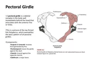
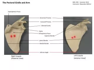
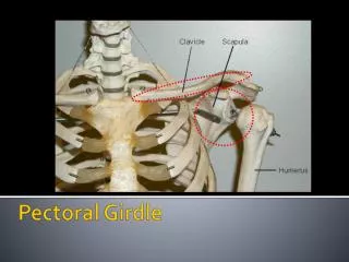
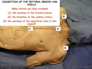
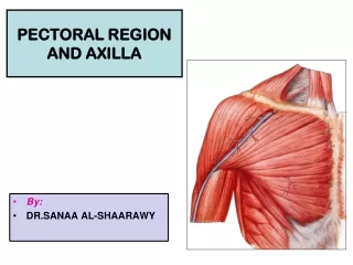
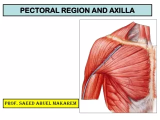
Muscles of the pectoral region • Each pectoral region contains the pectoralis major, pectoralis minor, and subclavius muscles • All originate from the anterior thoracic wall and insert into bones of the upper limb.
Pectoralis major • The pectoralis major muscle is the largest and most superficial of the pectoral region muscles • It directly underlies the breast and is separated from it only by deep fascia and the loose connective tissue of the retromammary space. • Pectoralis major has a broad origin that includes the anterior surfaces of the medial half of the clavicle, the sternum, and related costal cartilages (upper six ) • The muscle fibers converge to form a flat tendon, which inserts into the proximal end of the humerus (Lateral lip of bicipital groove ). • Pectoralis major adducts, flexes, and medially rotates the arm. • InnervationMedial and lateral pectoral nerves
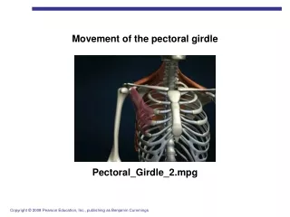
Subclavius and pectoralis minor • The subclavius and pectoralis minor muscles underlie pectoralis major • subclavius is small and passes laterally from the anterior and medial part of rib I to the inferior surface of the clavicle • pectoralis minor passes from the anterior surfaces of ribs III to V to the coracoid process of the scapula • Both subclavius and pectoralis minor pull the tip of the shoulder inferiorly, Subclavius Pulls clavicle medially and downward to stabilize sternoclavicular joint. • A continuous layer of deep fascia, clavipectoral fascia, encloses subclavius and pectoralis minor and attaches to the clavicle above and to the floor of the axilla below. • InnervationNerve to sub-clavius, Medial pectoral nerves