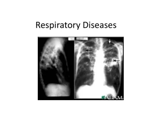
RESPIRATORY DISEASES
RESPIRATORY DISEASES . Hypoxemia/Hypercapnia. Hypoxemia =/= hypoxia Hypoxia = decr’d oxygen at tissues Hypoxemia = low PaO 2 V/Q Ratio ventilation rate(V) compared to perfusion rate(Q) of blood through lung capillaries at alveoli Normal (healthy) ratio = 0.8.

RESPIRATORY DISEASES
E N D
Presentation Transcript
Hypoxemia/Hypercapnia • Hypoxemia =/= hypoxia • Hypoxia = decr’d oxygen at tissues • Hypoxemia = low PaO2 • V/Q • Ratio ventilation rate(V) compared to perfusion rate(Q) of blood through lung capillaries at alveoli • Normal (healthy) ratio = 0.8
PaCO2dependent onVA (alveolar ventilation rate) • If CO2 removal =/= CO2 prod'n, PaCO2 will increase • If decr’d VA (hypoventilation) incr’d PaCO2 (hypercapnia) • Ventilation not adequate to meet metabolic demands (cells producing more CO2 than lungs can get rid of)
If CO2 removed faster than produced, PaCO2 will decrease • If incr’d VA (hyperventilation), decr’d PaCO2 (hypocapnia) • Ventilation exceeds metabolic demands • Occurs with • Anxiety • Head injury • Insufficient O2 in blood (Why would you now breathe faster?) • Remember acid/base imbalances: what happens to acid/base balance if you breathe too little? What compound is increased in the blood? Why is this bad? What would your body do to compensate?
So, with respect to V/Q Ratio: • If V/Q < 0.8, body has “wasted perfusion” (physiologic shunt) • Blood “shunted” past alveoli w/out adequate gas exchange • Due to impaired ventilation • PaO2 decr’d • PaCO2 incr’d
Shunting - alveolar spaces nonfunctional • If extreme physiologic shunt (if alveoli collapsed, edematous, filled w/ exudates), get extreme V/Q mismatch (V=0) • Can cause: • Severe hypoxemia • Administration oxygen does not correct
If V/Q > 0.8, called dead-space disease • Q = 0 (again, extreme V/Q mismatch; now due to no perfusion) • Ventilation not effected physiologically, BUT see incr’d work of breathing • Also hypercapnia, hypoxemia • Clinical - variable response • Hypoxemia if PaO2 = 40-50 mm Hg • Impairment of • Brain – headache, confusion, unconsciousness • Heart – tachycardia, dysrhythmias, incr’d bp • Hypercapnia • CNS depressed (headache coma) • Body fluids become acidotic
Pathologies vent’n/ perfusion imbalance • Heart disease • Incr’d pressures in lung vasculature (pulmonary hypertension) • W/ valve disorders, CHF, etc. • pulmonary arteriosclerosis decr’d blood volume at lung (hypoperfusion) • So decr’d Q and V/Q mismatch • Note: not the same as incr’d perfusion • Actually, more similar to ischemia to lung tissues
Pulmonary embolism hypoperfusion • Material in pulmonary circulation “plugged” vessel • Thromboembolism most common • Often w/ thrombi from lower extremities • Same risk factors as w/ systemic thromboembolism (smoking, hyperlipidemia, hypertension, etc.) • Most common cause of acute pulmonary disease in US • ½ deaths occur w/in 2 hrs of embolism in pulmonary circulation • Pathophysiology • V/Q mismatch (decr’d Q) incr’d PaCO2 and decr’d PaO2 • Also pulmonary hypertension may result (Why?) and/or decr’d CO
Pulmonary embolism – cont’d • Clinical – severity varies • Massive • shock, chest pain, tachypnea, tachycardia • With lung infarct, see pleural pain, pleural effusion, dyspnea • What does the term infarct mean? • Treatment • Prevention • Eliminate risks associated w/ thrombus formation (stop smoking, etc.) • Anticoagulants • Prevent clot formation
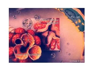
Pulmonary edema • Excess fluid in lung, so decr’d ventilation • Lung usually kept “dry” by 3 mechanisms: 1) lymph drainage 2) capillary exchange 3) surfactant lining the alveoli • Predisposing factors to pulmonary edema related to 3 • Heart disease • LV failure incr’d pressures in L heart incr’d pressures in respiratory (gas exchange) capillaries • Fluid gets forced out of capillaries alveoli and between lung cells • So decr’d gas exchange ability and decr’d lung compliance • Lymph drainage compensates for awhile
Capillary injury • Injury incr’s capillary permeability fluid forced more easily out of capillaries into alveoli and between cells (as above) • Decr’s compliance and gas exchange • Injury to cap’s may be due to chemical, physical lung injury • Obstruction of lymph system • If lymph vessels or nodes blocked little/no drainage of ISF (so builds up between lung cells and into alveoli) • Again decr’d compliance and gas exchange • Clinical • Dyspnea, hypoxemia, incr’d work of breathing • Treatment • Depends on cause
Obstructive Respiratory Diseases • incr’d resistance to air flow and decr’d vent’n • Due to obstruction • In lumen of airway (ex: incr’d secretions w/ asthma) • In airway wall (ex: inflamm’n at bronchial epithelium w/ asthma, chronic bronchitis) • In structures surrounding smooth muscle (ex: contraction bronchial smooth muscle w/ asthma) • Most common obstructive diseases: chronic bronchitis, emphysema, asthma • Difficult expiration
Chronic bronchitis • Inflammation of bronchi • Most common obstructive disease • Caused by • Irritants (dominant – cigarette smoke) • Also infection • Chronic bronchitis leads to: • Increased mucus secretion • AND thickening of mucus
AND thickening of bronchial mucosa layer (w/ hypertrophy & hyperplasia of bronchial epithelium) • Body’s compensations for chronic irritation • Tries to guard cells that make up airways from irritation • Get diffuse obstruction • Lung defense mechanisms compromised • Cilia impaired • Thickened mucus can’t get rid of invaders • Find incr’d acute respiratory infections
Pathophysiology • Airways collapse w/ expiration • V/Q mismatch • PaCO2 incr’d (hypercapnia); hypoxemia • Clinical • Wheezing, shortness of breath • Exercise tolerance decr’d • Productive cough (sputum coughed up) • Incr’d risk respiratory infections • Treatment • Prevention -- STOP SMOKING • Bronchodilators open lumen • Expectorants decr mucus thickness
Emphysema • destruction of alveolar walls, so decr’d elastic recoil of alveoli • Causes decr’d ability to expire • Note: obstruction not due to physical substance causing blockage; rather obstruction to gas exchange, air movement due to change in lung tissue • Genetic predisposition • Pathophysiology • Destruction of alveolar septa (shared cell membr between 2 alveoli) • Large air spaces develop (bullae) • Airways enlarge
Clinical • Dyspnea on exertion developing to dyspnea at rest • No cough; little sputum (REMEMBER: no incr’d mucus) • Tachypnea (incr’d rate of breathing) • Treatment - as per chronic bronchitis • Together, chronic bronchitis + emphysema = Chronic Obstructive Pulmonary Disease (COPD) or Chronic Airway Obstruction (CAO)
Asthma • Inflammatory disease • Reversible • Unlike chronic bronchitis + emphysema • Bronchospasm + mucus hypersecretion + swelling w/ inflamm’n • Bronchospasm = prolonged contraction bronchial smooth muscle • Obstruction of air flow
Due to: • Hyperactive immune response to allergens • Prod incr’d amounts of IgE • Bind mast cells in airways • Much histamine released • Hyperactivated immune, inflammatory responses • Also, bronchospasm, incr’d mucus and edema w/ incr’d capillary permeability
Due to (cont’d) • Neural dysfunction with dysfunctional autonomic nervous system response • Disrupted or hyperactive irritant receptors (?) • Bronchospasm, perhaps also due to mast cell involvement (so all histamine effects noted above) • Common in children • Approx 50% of all asthma cases • Remission common (as adults) when asthma begins in childhood
Pathophysiology • Vascular congestion, edema • Formation of thick mucus impaired ciliary function • Incr’d work of breathing • Hyperventilation • Clinical • Wheezing, nonproductive cough, tachycardia, mucus formation • Treatment • Eliminate cause of attack • Drugs to reverse bronchospasm
Restrictive respiratory disease (extrapulmonary) • Lung tissue is normal, other disorders affect ventilation • Chest wall restrictions • Work of breathing incr’s and ventilation decr’s • Hypoventilation, hypercapnia, hypoxemia • Impaired lung defenses • “Stagnant” air (doesn’t move out of body as it should) • If contains microbes or invaders, incr’d risk of infecting airways -- more time to grow, replicate
Due to • Chest wall deformities • Fat overlaying chest muscles in very obese patients • Neuromuscular diseases (ex: polio, muscular dystrophy, others) • Dyspnea • Patients more susceptible to lower resp tract infections • Over time, can respiratory failure
Intrinsic restrictions – • Acute or chronic • Chronic • Chronic Intrinsic Restrictive Lung Disease (CIRLD) • Excessive fibrous/connective tissue deposits in lung • Lung injury scar tissue formation • Lung stiffness, so compliance decr’s • Due to: • Irritant inhalation, infection, autoimmune dysfunction • Leads to • Decr’d ventilation (harder to breathe) • Hypoxemia • V/Q mismatch
Acute restrictions • ARDS - Adult Respiratory Distress Syndrome • Due to injury to lung • Direct: inhaling toxic gases, trauma • Indirect: systemic disorder incr’d chemical mediators of infection (thromboxanes, etc.) • (Biochem’s similar to prostaglandins; patients either release in too high concentrations, or lung cells too sensitive to them) • Pathophysiology: • Acute lung inflammation • Severe pulmonary edema • Diffuse alveolo-capillary injury • Severe pulmonary edema, hemorrhage
Pathophysiology – cont’d • Get decr’d compliance, decr’d alveolar ventilation, incr’d pulmonary vasoconstriction • Fibrosis within 7 days of injury ARDS • Clinical • Rapid, shallow breathing • Marked dyspnea • Hypoxemia unrelieved by oxygen administration • Fatal in ~ 70% of cases • Treatment • Mechanical vent’n to incr available oxygen • Sedation to decr oxygen consumption • Increase C.O., give diuretics to relieve edema
Lung Infections • When lung defenses decreased • Pneumonia • By bacteria or virus • Common bacteria = strep pneumoniae • Causes ~70% of all pneumonia • Pathophysiology • Pathogens multiply in lung • Overall, due to decr’d immune response in lung • Toxins released from microbes • Bronchial mucosa becomes damaged
Pathophysiology – cont’d • Inflammation/edema results • Exudate found in alveoli • V/Q mismatch (decreased V) • Clinical • Infection chills/fever/malaise • Chest edema cough, pleural pain • Dyspnea • Treatment • Antibiotics – for bacterial infection • Mechanical ventilation
Atelectasis = collapse of lung (alveoli) • Two types • Compression • External pressure pushes air out of alveoli • Alveoli can’t re-expand (because of increased pressure still on lung)
Absorption • Occurs w/ obstruction, when no expiration/inspiration • “Old” air absorbed from alveolus over time, not replaced • So alveoli collapse • Seen post-operatively • Anesthetics cause incr’d mucus production obstruction • Clinical • Dyspnea, cough, fever
Pleura, pleural space affected • Pleural effusion = fluid (blood, lymph) in pleural space • Can cause collapse of lung tissue • Due to incr’d pressure of fluid pressing on alveoli • Hemothorax = bleeding into pleural space • Empyema = infected pleural effusion • Seen w/ lymph blockage • Pneumothorax = air/gas in pleural space • Negative pressure in pleural space destroyed • Pressure differential nec for proper pressures, recoil • Due to trauma, secondary to thoracic surgery
Lung Cancers • Bronchogenic carcinoma • Malignant tumors of mucous membranes • Larger bronchi • ~90% of all primary lung cancers • Epidemic in U.S. • ~200,000 new cases per year • Most common of all primary tumors; most frequent cause of cancer death
Most common cause of bronchogenic carcinoma: • Cigarette smoking • Heavy smokers ~25X greater risk than nonsmokers • Other causes • Environmental, occupational (breathing in noxious/traumatizing agents, such as asbestos)
Classification by histological type; each type treated differently • Non-Small Cell Lung Cancer • Treated surgically • Squamous cell carcinoma – most common • Centrally located • Remains localized • Metastasis relatively late • Associated w/ smoking
Non-small cell cancers – cont’d • Adenocarcinoma • Tumors arise in periphery of lung • Most common in women • One type = bronchioalveolar cell carcinoma • Slow growing • Weak association w/ cigarette smoking • Low survival rate • Asymptomatic w/ early metastasis
Small Cell Lung Cancer • Treatment by chemotherapy, radiation • Strongest association w/ cigarette smoking • 20-25% of all bronchogenic carcinomas • Oat cell carcinoma • Cells compressed • Rapid growth, early metastasis • Poor prognosis (<5% alive in 2 yrs) • Stages common to epithelial cancers • Irritation hyperplasia, metaplasia, neoplasia, etc.