Respiratory Diseases
Respiratory Diseases. J. Matthew Velkey m att.velkey@duke.edu 454A Davison, Duke South, Green Zone. Atelectasis (collapse). Resorption Obstruction (e.g. infection or inflammation or mucous plug in CF patients): air downstream of blockage is slowly resorbed Compression

Respiratory Diseases
E N D
Presentation Transcript
Respiratory Diseases J. Matthew Velkey matt.velkey@duke.edu 454A Davison, Duke South, Green Zone
Atelectasis (collapse) • Resorption • Obstruction (e.g. infection or inflammation or mucous plug in CF patients): air downstream of blockage is slowly resorbed • Compression • Pneumothorax (air leak into pleural cavity) • Hemothorax or other fluid • Contraction • Localized fibrosis causes mechanical contraction and collapse of lung tissue
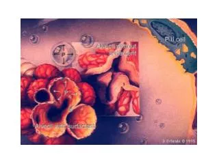
ARDS: Acute Respiratory Distress Syndrome • Occurs in the setting of sepsis, severe trauma (smoke inhalation, drowning, etc.), and/or pulmonary infections • Imbalance in pro- and anti-inflammatory mediators • NF-kB induces mphages to release TNF, Il-8, & Il-1 which recruits and activates neutrophils • Activated neutrophils release factors that destroy tissue and recruit MORE inflammatory cells (positive feedback loop) • Results in diffuse damage to capillary and alveolar epithelium • Effusion of blood and plasma due to capillary leakage • Loss of surfactant due to type II pneumocyte damage
Obstructive vs. Restrictive Pulmonary Diseases • Obstructive (e.g. emphysema, asthma, bronchitis) • Limitation of airflow due to partial or complete airway obstruction • Total lung capacity and forced vital capacity (FVC) are NORMAL, but Forced Expiratory Volume (FEV) is decreased (i.e. FEV:FVC ratio REDUCED) • Restrictive (e.g. fibrosis, asbestosis, sarcoidosis) • Reduced expansion due to lung fibrosis • FVC and FEV reduced proportionately
Emphysema • Chronic obstructive airway disease characterized by permanent enlargement of airways distal to bronchioles • Imbalance of protease/antiprotease activity and/or excess of reactive oxygen species activity • Excessive activation of neutrophil and macrophage elastase associated with chronic smoking • Hereditary α1 anti-trypsin deficiency • Dyspnea and hyperventilation but with adequate gas exchange (until very late in disease), aka “pink puffers”
Chronic Bronchitis • Aka, “small airway disease” • Persistent productive cough for at least 3 months in at least 3 consecutive years • Hypersecretion of mucus, hypertrophy of mucous glands • Infiltrates of neutrophils, macrophages, and CD8+ T cells (IL-13 release stimulates mucus secretion) • Mucus plugging may completely occlude bronchiolar airways (bronchiolitis obliterans) and often results in opportunistic infections • Associated with hypoxia, cyanosis, and obesity, aka “blue bloaters”
COPDChronic Obstructive Pulmonary Disease • Emphysema AND chronic bronchiolitis usually co-exist and are clinically grouped as “COPD” • Affects ~10% of US population, 4th leading cause of death • Irreversible airflow obstruction (vs. asthma in which obstruction is reversible) and (usually) pulmonary hypertension
Asthma • Intermittent, reversible airway obstruction • Chronic eosinophilic bronchial inflammation • Bronchial smooth muscle hypertrophy and hyperreactivity • Usually extrinsic or “atopic” (immune hypersensitivity) but can be intrinsic or “non-atopic” (triggered by non-immune stimuli) –regardless of type clinical picture is the same
Asthma pathogenesis • Sensitization phase: antigens induce TH2 cells • TH2 cells secrete factors that stimulate IgE production by Plasma Cells • IgE binds to receptors on Mast Cells • Mast Cells produce factors that recruit Eosinophils to tissue
Asthma pathogenesis Reactive phases: Immediate: antigen binding to IgE on Mast Cells stimulates degranulation, releasing factors (e.g. leukotrienes, prostaglandin, histamine) that cause bronchoconstriction, edema, and mucus secretion AND recruit more inflammatory cells (particularly eosinophils). Late: Eosinophilsdegranulate, releasing factors that damage bronchial tissue
Bronchiectasis • Permanent dilation of bronchi and bronchioles caused by destruction of tissue secondary to chronic infections • Bronchial obstruction: e.g. mucus plugs in CF patients, chronic bronchitis, asthma, or Kartagener syndrome facilitate infections • Immunodeficiency (HIV, transplant). • Usually affects lower lobes • Characterized by markedly dilated bronchi and distal airways and intense acute inflammation within bronchi and bronchioles
Diffuse Interstitial (Restrictive) Lung Diseases • Diffuse, chronic fibrosis of pulmonary connective tissue (i.e. the “interstitium”) surrounding alveoli • Stiffens and thickens lung tissue thereby reducing the airspace (FVC) AND the expiratory flow rate (FEV1) proportionately • Activated macrophages are the key player: • Damage alveoli and recruit neutrophils • Secrete factors that induce fibrosis • Damaged alveoli also induce fibrosis Prototypical exemplars are Idiopathic Pulmonary Fibrosis, Pneumoconioses, and Sarcoidosis
Idiopathic Pulmonary Fibrosis • Unknown etiology (hence idiopathic) • M > F, mostly > 60 years old • Presents with dyspnea and non-productive cough and progresses relentlessly • Characterized by patchy interstitial fibrosis • Eventually collapses alveoli and large, cystic spaces form giving the lung a honeycomb appearance
Pneumoconioses • Chronic fibrosing disease due to chronic exposure to particulates • Pulmonary macrophages play central role by promoting inflammation, alveolar damage, and fibrosis • Can progress to pulmonary dysfunction, pulmonary hypertension, and right-sided CHF • Examples include anthracosis (black lung), silicosis, and asbesosis
Anthracosis(black lung) • Chronic exposure to coal dust • Tends to affect upper lobes first
Silicosis • Chronic exposure to silica (sandblasting, drywall compound) • Characterized by silicotic nodules • Tends to affect upper lobes first
Asbestosis • Chronic exposure to asbestos • Characterized by asbestos bodies in the lung and fibrotic plaques in the parietal pleura • Tends to affect middle and lower lobes
Sarcoidosis • Multisystemic disease of unknown etiology • Higher incidence in Scandinavian and African-American population, usually <40 yo • Lung tissuecontains high levels of CD4+ TH1 cells and associated cytokines (IFNγ & IL-2) • Characterized by formation of non-caseating granulomas and interstitial fibrosis
Hypersensitivity Pneumonitis • Hypersensitivity reaction similar to asthma, however damage occurs in the ALVEOLI, so presents as a RESTRICTIVE lung disease rather than obstructive • Characterized by patchy mononuclear cell infiltrates and increased numbers of CD4+ and CD8+ T cells • Interstitial non-caseating granulomas may also be present
Pulmonary Hypertension • Often occurs with chronic obstructive or interstitial lung disease or secondary to cardiovascular disease • Can also be caused by recurrent embolization or mitral valve stenosis (blood can’t flow into LV as well so LA pressure increases and backs up into lungs • Often backs up blood into RV and may lead to right sided CHF • Vascular alterations in pulmonary arteries include: • Atherosclerosis in distributing elastic arteries • Medial and intimal thickening in muscular arteries • Medial thickening and IEL disruption in small arteries, sometimes completely occluding lumen • Plexogenic pulmonary arteriopathy: tufts of capillaries spanning small arteries
Pneumonia • Infection (usually bacterial) of the lung • Cause of one-sixth of all deaths • Two general patterns: • Lobar: entire lobes are involved • Bronchopneumonia: foci of inflammation corresponding to bronchial tree, usually bilateral and basal • Regardless of pattern, key to treatment is knowing what bug is causing the infection, the usual suspects are: • Streptococcus pneumoniae(particularly w/ CHF, COPD, or Diabetes) • Haemophilusinfluenzae(often in children or adults w/ chronic pulmonary disease) • Moraxella catarrhalis (often in elderly or adults with COPD) • Staphylococcus aureus(often after respiratory illness or associated with staph endocarditis) • Klebsiellapneumoniae(often in malnourished individuals) • Pseudomonas aeruginosa(often in hospital setting w/ neutropenic patients) • Legionella pneumophila(often in transplant patients or those with chronic heart, renal, or blood disease)
Pneumonia: clinical course • Congestion: affected lobes are congested, red, and boggy • Red hepatization: tissue is red and firm with a liver-like consistency; alveoli are packed with red blood cells, fibrin, and neutrophils • Gray hepatization: lung is gray due to lysis and ingestion of red blood cells and still firm and liver-like due to persistence of fibrinopurulent exudate within alveoli • Resolution: fibrinopurulent exudate mostly resorbed by macrophages but can also organize into fibrous scar Possible complications include: Pleuritis: fibrinopurulent reaction in pleura may result in adhesions Abcessformation and cavitation (loss of alveoli) Empyema: accumulation of suppurative exudate within pleural cavity Fibrosis Bacteremic dissemination leading to meningitis, arthritis, or endocarditis
Tuberculosis Chronic granulomatous disease caused by Mycobacterium tuberculosis, usually affecting the lung, but can affect any organ via hematogenous spread • Initial exposure induces immune reaction that usually quells the infection and confers resistance, but foci of infection can harbor viable pathogens for years that can be reactivated in states of diminished immunity • Primary exposure and/or secondary “reactivation” in immune compromised can result in massive, life-threatening systemic infections
Tuberculosis features Hematogenous dissemination or “miliary” tuberculosis (so-called because of the millet seed appearance of infection foci) Granulomas (often caseating but not always) ringed by epithelioid macrophages and giant cells
Lung Cancers • #1 cause of cancer-related deaths • Peak incidence in 50s and 60s • At diagnosis, over 50% of patients already have metastatic disease. • Overall 5yr survival rate is ~15% • Often associated with paraneoplastic endocrine syndromes due to hypersecretion of various hormones such as PTH or ACTH 4 general types: • Squamous cell carcinoma: more common in men, squamous metaplasia within major bronchi • Adenocarcinoma: usually peripherally located, arise from mucous glands • Large-cell carcinoma: undifferentiated, epithelial tumors (most likely started out as either squamous or adeno- but have de-differentiated such that they are no longer recognizable as such) • Small-cell carcinoma: pale, gray, central masses; high mitotic index; likely derived from neuroendocrine cells; worst prognosis (5. Bronchial carcinoid: arise from Kulchinsky neuroendocrine cells, often resectable and curable)