Case 11
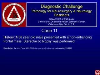
Diagnostic Challenge Pathology for Neurosurgery & Neurology Residents Department of Pathology University of Oklahoma Health Sciences Center, Oklahoma City, OK, U.S.A. Case 11 History: A 58 year-old male presented with a non-enhancing frontal mass. Stereotactic biopsy was performed.

Case 11
E N D
Presentation Transcript
Diagnostic ChallengePathology for Neurosurgery & Neurology ResidentsDepartment of PathologyUniversity of Oklahoma Health Sciences Center,Oklahoma City, OK, U.S.A. Case 11 History: A 58 year-old male presented with a non-enhancing frontal mass. Stereotactic biopsy was performed. Contributor: Kar-Ming Fung, M.D., Ph.D., karming-fung@ouhsc.edu Last updated: 1/9/2008
Cytologic Preparation Cytologic Preparation A B
Frozen Section Frozen Section C D
Paraffin Section Paraffin Section E F
Ki67 p53 G H
Diagnosis: At least diffuse astrocytoma, at least WHO II. • Discussion: • In the cytologic preparation, the atypical cells with elongated cytoplasmic processes (white arrow below) and mucoid material (black arrow) are diagnostic for a glial neoplasm. (Panel A and B) • The overall cellularity is low and mitotic figures are not readily seen. (Panel C to F) Ki67 labeling is low. (Panel G) • These features are diagnostic for a low-grade (WHO II) astrocytoma. What is unusual is that this type of tumor is not common in patients over 50 years of age. Although there is no enhancement, the presence of high grade tumor that are not included in this stereotactic biopsy is possible. • P53 is positive in this case but this feature does not help to confirm the diagnosis. This tumor is also positive for glial fibrillary acidic protein (GFAP) (not shown.