ICP & Head Trauma
ICP & Head Trauma. Sophia R. Smith, MD WRAMC November 2, 2005. Introduction. Head injuries are one of the most common causes of disability and death in children.

ICP & Head Trauma
E N D
Presentation Transcript
ICP & Head Trauma Sophia R. Smith, MD WRAMC November 2, 2005
Introduction • Head injuries are one of the most common causes of disability and death in children. • The Centers for Disease Control and Prevention (CDC) estimates that more than 10,000 children become disabled from a brain injury each year. • Head injuries can be defined as mild as a bump to severe in nature.
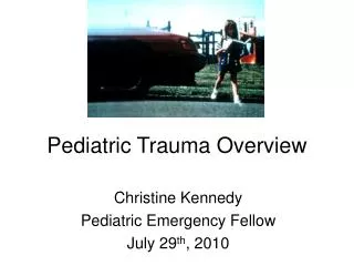
Prevalence of Pediatric Trauma • Trauma is the leading cause of death in infants and children • Trauma is the cause of 50% of deaths in people between 5 and 34 years of age • Motor vehicle related accidents account for 50% of pediatric trauma cases • $16 billion is spent annually caring for injuries to children less than 16 years of age
Primary Brain Injury Results from what has occurred to the brain at the time of the injury Secondary Brain Injury Physiologic and biochemical events which follow the primary injury Traumatic Brain Injury
Factors that Effect Secondary Brain Injuries Blood Pressure Oxygenation Temperature Control of Blood Glucose Fluid Volume Status Increased Intracranial Pressure
SOME of the SECONDARY EVENTS IN TRAUMATIC BRAIN INJURY diffuse axonal injury inflammation BBB disruption apoptosis necrosis edema formation Brain trauma ischemia energy failure cytokines Eicosanoids endocannabinoids Calcium polyamines Acetyl Choline ROS Shohami, 2000 Green – pathophysiological processes; Yellow – various mediators
Anatomy of the cranium • There are various brain contents that are localized within a rigid structure. • Cranium • The cranial vault contents include: • The brain • The cerebral spinal fluid • The cerebral blood
Cerebral Spinal Fluid • CSF • 150 cc in adults at all times • Children slightly less • Produced by choroid plexus – 20 cc/hr • CSF is absorbed into venous system at the subarachnoid villi
Cerebral blood and brain • Cerebral blood • Sum of blood in capillaries, veins, and arteries • Brain • 80% of the total intracranial volume • All of these contents are maintained @ a balanced pressure referred to as intracranial pressure (ICP)
Monro-KellieDoctrine • The ICP within the skull is directly related to the volume of the contents. • Defined as the Monro-Kellie Doctrine • This doctrine states that any increase in volume of the contents within the brain must be met with a decrease in the other cranial contents.
Monro-Kellie Doctrine Vintracranialvault=Vbrain+Vblood +Vcsf
Cerebral Blood Flow • CBF is directly linked to the metabolic requirements of the brain. • As the brain metabolic activity increases, CBF increases • Vasodilatation of cerebral vessels • Increase in cerebral blood volume • Consequent increase in ICP
Cerebral blood flow • CBF maintained when MAP range is 50mmHg to 150mmHg • Cerebral auto regulation • As BP increase baroreceptors sense event and cerebral arteries vasoconstrict CBF maintained with a CBV decrease • As BP decrease cerebral arteries dilate to increase flow CBV increase
Auto regulation • This process is lost in pathological states • Esp. Head trauma • CBF decreases linearly to MAP below range • Results is ischemia (strokes) to brain regions • CBF increases linearly to MAP above auto regulation range • HTN encephalopathy as CBV and ICP increase
Mediators of CBF • Local and global mediators of CBF and metabolism are important. • Hypoxia and pHare most important • As local paO2 decreases, CBF increases • CBF is affected by pH (and its surrogate pCO2)
Blood: Cerebral Blood Flow • The brain has the ability to control its blood supply to match its metabolic requirements • Chemical or metabolic byproducts of cerebral metabolism can alter blood vessel caliber and behavior
Studies of hyperventilation & ICP • This relationship has been well studied as a therapeutic option in particular intentional hyperventilation to lower cerebral blood flow and thus intracranial pressure. • No longer a practice • Modest hyperventilation
On call • So, you are in the ER on your first night of call and the next thing you know you get your very first trauma patient. • How do you evaluate?
Glascow Coma Scale Eye Opening Spontaneous 4 To Voice 3 To Pain 2 None 1Best Verbal Oriented 5 Confused 4 Inappropriate Words 3 Incomprehensible Sounds 2 None 1Best Motor Obeys Commands 6 Localizes Pain 5 Withdraws to Pain 4 Flexion to Pain 3 Extension to Pain 2 None 1
Severe TBI • Indications for Intubation • GCS< 8 • Fall in GCS of 3 • Unequal pupils • Inadequate respiratory effort or significant lung/chest injury • Loss of gag • apnea
Treatment • Intubation. • Pretreatment with lidocaine 1 mg/kg IV may prevent rise in intracranial pressure (ICP).
Treatment • Hyperventilation • to maintain PO2 >90 torrs, PCO2 30 to 32 torrs. • Hyperventilation may actually increase ischemia in at risk brain tissue if PCO2 <25 torr by causing excessive vasoconstriction and has fallen out of favor. Prophylactic hyperventilation for those without increased ICP is contraindicated and worsens outcomes. • PEEP relatively contraindicated because reduces cerebral blood flow.
Maintain normal cardiac output. • If hypotensive from other cause such as multi-trauma, treat shock as usual. • Normal saline is preferred over LR since LR is slightly hypotonic. • Hypertonic saline (3% or 7.5%) can be used. Especially if you see ICP changes.
Maintain normal cardiac output. • If markedly hypertensive, consider labetalol or nitroprusside. • Avoid lowering the blood pressure unless diastolic blood pressure is >120 mm Hg.
Diuresis • Mannitol 1 g/kg IV over 20 minutes induces osmotic diuresis. • Avoid if hypotensive or have CHF/renal failure. • Some suggest furosemide (Lasix and others). • Avoid if hypotensive.
ICP Precautions • Elevate head of bed 30 degrees. • Seizure prophylaxis: Phenytoin will reduce seizures in the first week after injury but does not change the overall outcome. • Steroids are ineffective in controlling ICP in the trauma setting.
Manipulation of CPP • CPP = MAP - ICP • Maintain adequate intravascular volume • CVP • Increase MAP • Utilize alpha agonist--dopamine, phenylephrine, norepinephrine • What is appropriate goal for children?
CPP for children • Aim for a CPP of >60 mmHg • by maintaining an adequate MAP and control of ICP • MAP – ICP = CPP • Minimizing the morbidity of TBI in children
Additional therapies • Prevent hyperglycemia: exacerbates ischemic cerebral damage • Attention to electrolyte status. These patients are prone to electrolyte abnormalities due to osmotic diuresis, cerebral salt losing states, SIADH and diabetes insipidus
Manipulation of ICP Blood • Decrease cerebral metabolic demand • sedation, analgesia, barbiturates • avoid hyperthermia • avoid seizures • Hyperventilation • decreases blood flow to brain • only acutely for impending herniation • Mannitol
Manipulation of ICP Brain • Mannitol • dehydrate the brain, not the patient! • monitor osmolality • Hypertonic saline • Decompressive craniectomy
ICP Monitoring • ICU patients who have sustained head trauma, brain hemorrhage, brain surgery, or conditions in which the brain may swell might require intracranial pressure monitoring. • The purpose of ICP monitoring is to continuously measure the pressure surrounding the brain.
Why Monitor? • Detect “events” • Manage intracranial pressure • Manage cerebral perfusion pressure
How? • Ventriculostomy • Intraparenchymal fiberoptic catheter • Subarachnoid monitor • Useful adjuncts: • Arterial line • Central venous line • Foley catheter
Manipulation of ICP CSF • External drainage • therapeutic as well as diagnostic • technical issues • infectious issues
What to do with the information... • Goal: adequate oxygen delivery to maintain the metabolic needs of the brain. • Intracranial pressure <20 • Cerebral perfusion pressure >50-70 mm Hg CPP=MAP-ICP
Indications for ICP monitoring • Glasgow coma scale <8 • Clinical or radiographic evidence of increased ICP • Post-surgical removal of intracranial hematoma • Less severe brain injury in the setting which requires deep sedation or anesthesia
Other monitoring devices • CT Scan • MRI • PET Scan • Jugular Venous Oxygen Saturation
Near-infrared Spectroscopy • Uses absorption characteristics of oxy Hgb, deoxy Hgb, and [o] cyt aa3 • Uses the ability to penetrate the superficial brain • Therefore the state of oxygenation can be determined. • Good assessment of cerebral oxygenation
Transcranial Doppler US • TCD is a noninvasive technique used to determine cerebral blood velocity in large intracranial arteries. • Assessment of • Brain death • Reperfusion injury • Identify regions S/P TBI that are adversely effected
Cerebral Microdialysis • Measuring the partial pressure of oxygen of brain parenchyma and metabolites using microdialysis • Electrode in vulnerable brain region measures O2 concentration • Measures also local brain metabolism