Download
1 / 28
280 likes | 582 Vues
Management of Severe Head Trauma in A&E. Dr. David Tran A&E department FVHospital 17 mars 2010. Management of head injury. Primary survey: ensure that airways, breathing, circulation and cervical spine are secure. Assessment of mental state (glasgow score adapted to the age)

E N D
Management of Severe Head Trauma in A&E Dr. David Tran A&E department FVHospital 17 mars 2010
Management of head injury • Primary survey: ensure that airways, breathing, circulation and cervical spine are secure. • Assessment of mental state (glasgow score adapted to the age) • Alert (A), responds to voive (V), Responds to pain (P), unresponsive (U)
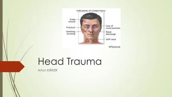
Perform secondary survey • Neck & cervical spine (tenderness, muscle spasm) • Head: scalp hematoma, laceration, swelling, tenderness… • Eyes: pupils size, equality, reactivity • Ears: otorrhage, hemotympan • Nose, mouth, facial fractures • Motor function: limbs, reflexes, lateralised weakness, Babinski’s sign.
Seriousness of head trauma • Glasgow Score < or = 8 (severe head trauma) • Glasgow score 9-12 (moderate head trauma) • Glasgow score > or = 13 (mild head trauma)
Do not forget neck protection • Severe head trauma are frequently associated with neck injuries. • Those injuries often concern the cervico-occipital region (C1/C2) • Neck collar has to be put immediately at arrival in A&E and will be removed only after imaging.
General management • IV line, use infusion of isotonic solutions* (NaCl 0.9% is the most adapted) • Intubation: all patients with severe head trauma (Glasgow score < or =8) have to be intubated. • Crash induction is the gold standard for management of airways. * Avoid hypotonic solutions like G5% or Ringer Lactate
Induction / sedation • Crash induction: • Etomidate 0.3mg/Kg & • Suxamethonium 1mg/Kg • Orotracheal intubation (becareful of the neck) • Immediate sedation with Hypnovel / Fentanyl IV is very important to control intracranial pressure (continuous infusion following protocole sedation in SMUR: 10 amp. Hypnovel /1 amp. Fentanyl)
Interest of early sedation in case of severe head trauma • Control agitation of the patient • Control of analgesia • Avoid or decrease intra-cranial hypertension. • Adaptation to mechanical ventilation
Monitoring head trauma in A&E • Non invasive blood pressure/15 min • SpO2 • Pulse rate on scope • PCO2 (if not available, blood gaz during mechanical ventilation)
Intracranial pressure monitoring (PPC= MAP-ICP) • Each time there is a severe traumatic brain injury (with GCS 3-8) associated with CT scanner images of hematomas, contusions, swelling, herniation or compressed basal cisterns.
Head CT scanner abnormalities • Left Epidural hematoma • Effacement of left ventricle • Shift >10mm of the medium line • Signs of ICH
Head CT scanner abnormalities (2) • Right Sub-dural hematoma • Shift > 10mm of the medium line • Effacement of the right ventricle
Head CT scanner abnormalities (3) • Frontal contusions (hemorrhages) • Effacement of the cisterns and sub-arachnoid spaces
Head CT scanner abnormalities (4) Hyperdense lesion in R. frontal lobe = cerebral contusion Biconvexity in the left petrus temporal = HED
CT scanner abnormalities (5) Ventricles & sub-arachnoid spaces obliterated = ICH Sub-arachnoid hemorrhage
CT Scanner abnormalities (6) Sub-arachnoid hemorrhage in posterior fossa
Recommandations • Blood pressure should be monitored and hypotension avoided (Pas > 90mmHg) • Oxygenation should be monitored and hypoxemia avoided (Pa O2 > 60mmHg SpO2>90%) • Mannitol is effective for control of raised intra-cranial pressure (ICP)
Signs of intra-cranial hypertension (ICH) • Signs of transtentorial herniation / ICH: anisocoria, mydriasis, neurological lateral signs, seizures, bradycardia, hypertension, bradypnea. • Progressive neurological deterioration not attributable to extra-cranial causes. • Those signs are an indication for immediate use of bolus of Mannitol (up to 1mg/Kg/20 min.)
Use of Mannitol • Indication: Signs of intra cranial hypertension • Mannitol 20 or 25% (20g/100ml or 25g/100ml) • Bolus 0.25 to 1g/Kg/20 min. • Exp: body weight 60 kgs > 15g to 60g IV in 20 min. = 100 to 300ml Mannitol 20%
Administration of Mannitol • Mannitol is superior to Barbiturates for control of high ICP after TBI. • The osmotic effect of Mannitol is delayed for 15-30 min. while gradients are established between plasma & cells. • Its effects persist for about 90 min. to several hours.
Use of hypertonic Saline (HS) • Osmotic mobilization of water across blood brain-barrier. (Saline 7,2% or 10%) • HS as a bolus infusion could be an effective adjuvant to Mannitol to treat ICH. • Potential side effects: central pontic myelinolysis in patient with chronic hyponatremia. • More studies are required to determine the place of HS in the treatment of ICH.
Goals for management of severe head trauma • Any episode of hypotension or hypoxia increases head injury mortality. • Systolic blood pressure > 90mmHg (ideal = SBP 120mmHg & MAP 85mmHg ) • SpO2 > 90% (PaO2> 60mmHg)
Management of severe head trauma in A&E • Neck collar • Monitoring BP, pulse, SaO2 • IV line and fluid infusion (NaCl 0.9%) to restore systolic BP >90mmHg • Intubation (crash induction) and mechanical ventilation. • Immediate sedation after intubation (hypnovel/fentanyl)
Interest of early CT scanner • CT scanner has to be performed before transfert to neurosurgical center. • The time you spend to perform CT in FVH (<15min.) is time you save for the patient later. (timing in CR is probably longer)
Management of imaging • Head CT scanner without injection • Complete by images of cervico-occipital and cervical region. • Chest Xray and Pelvis Xray are systematic • Thorax, abdomen and dorso-lombar rachis CT scanner are requested according clinical signs.