Head trauma
Head trauma. Trauma department Hsinglin Lin. Introduction. Adequate oxygenation Maintenance of sufficient blood pressure Avoid secondary brain damage Early consultation BCT in hospital cannot treat. Consulting neurosurgeon 1.age and mechanism


Head trauma
E N D
Presentation Transcript
Head trauma Trauma department Hsinglin Lin
Introduction • Adequate oxygenation • Maintenance of sufficient blood pressure • Avoid secondary brain damage • Early consultation • BCT in hospital cannot treat
Consulting neurosurgeon 1.age and mechanism 2.Respiratory and cardiovascular status (BP) 3.Minineurologic ex., GCS( Motor response), pupillary reactions 4.associated injuries 5.Result of diagnostic studies (CT)
Eye Opening Response • Spontaneous--open with blinking at baseline 4 points • To verbal stimuli, command, speech 3 points • To pain only (not applied to face) 2 points • No response 1 point
Verbal Response • Oriented 5 points • Confused conversation, but able to answer questions 4 points • Inappropriate words 3 points • Incomprehensible speech 2 points • No response 1 point
Motor Response • Obeys commands for movement 6 points • Purposeful movement to painful stimulus 5 points • Withdraws in response to pain 4 points • Flexion in response to pain (decorticate posturing) 3 points • Extension response in response to pain (decerebrate posturing) 2 points • No response 1 point
Computed tomographic done in a patient has any of the following features: • The patient is eye opening only to pain or does not converse (Glasgow Coma Score 12/15 or less) • A deteriorating level of consciousness or progressive focal neurological signs • Confusion or drowsiness (Glasgow Coma Score 13 or 14/15) followed by failure to improve within at most four hours of clinical observation • Radiological/clinical evidence of a fracture, whatever the level of consciousness
New focal neurological signs which are not getting worse • Full consciousness (Glasgow Coma Score 15/15) with no fracture but other features, such as: • severe and persistent headache • nausea and vomiting • irritability or altered behaviour • a seizure
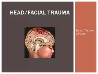
Anatomy • A: Scalp 1.skin, 2.connective tissure, 3.apponeurosis, 4.losse tissue, 5.pericranium. • B:Skull : cranial vault and base • C:Meninges: dura mater, arachnoid and pia mater. Most common injury: Middle meningeal a. in epidural space, Subdural space : bridge vein • D:Brain:cerebrum, cerebellum, brainstem
E: CSF • F: Tentorium:Oculomotor nerve runs along the edge of tentorium. Parasympathetic fibers lie on surface –dilation. Down and out with further compression. Uncal herniation: compression of the corticospinal tract in the midbrain - weakness of opposite side Kenohan’s notch syndrome: Same side
a) Subfalcial (cingulate) herniation ;b) uncal herniation ; c) downward (central, transtentorial) herniation ; d) external herniation ; e) tonsillar herniation.
Physiology • A: ICP: normal 10 mmHg, >20 mmHg: clear abnormal >40 mmHg: severe elevation • B: Cerebral perfusion pressure: <70 mmHg – poor outcome, CPP=MAP-ICP • C: cerebral blood flow: 50ml/100g of brain/min, <5ml/100g/min cell death, autoregulation : MAP 50-160 mmHg
Classication • A:Mechanism, 1.blunt:automobile collisions, fall, blunt assault, 2.penetrating: gunshot, stab w’d • B:Severity of injury: severe:GCS <8 ,moderate:9-13,mild:14-15. • C:Morphology and Injury: 1.Skull Fx, 2.Intracranial lesion.
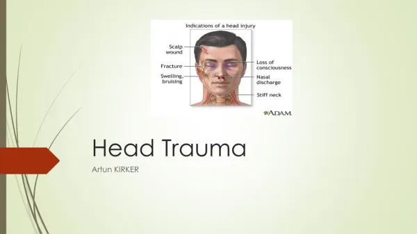
Skull fracture • Signs of Skull base fx: periobital ecchymosis (raccoon eyes), retroauricular ecchymosis (Battle’s sign), CSF leakage, 7th nerve palsy • Fragments depressed more than the thickness of the skull require surgical repair. • Skull Fx increases the likelihood of intracrainal hematoma.
Basilar skull fx are sometimes associated with CSF leakage from nose (rhinorrhea) or the ear (otorrhea). 7th nerve palsy.
Intracranial lesions • Focal lesions: • 1.EDH, often from middle meningeal a., relatively uncommon, treated early prognosis excellent, lucid interval, talk and die • 2.SDH, tearing of bridging vein, brain damage much sever and prognosis worse than EDH • 3.Contusion and intracerebral hematomas, associated SDH, frontal and temporal lobes • 4.diffuse injury- most common type of brain injury
Mild concussion consciousness preserved with noticeable degree of temporary neurologic dysfunction • Classic cerebral concussion-loss of consciousness , reversible, posttraumatic amnesia
Post-concussion syndrome- long-lasting neurologic deficits, include memory difficulties, dizziness, nausea, anosmia and depression. • Diffuse axonal injury- prolonged posttraumatic coma not due to mass lesion or ischemia insults. Decortication and decerebration with autonomic dysfunction.
Management of mild injury(GCS14-15) • CT – a history loss of consciousness, amnesia, or severe headaches. • observation at H for 12-24 hours • Skull X-ray – penetrating head injury, 1.linear or compression fx, 2.midline postion of pineal grand, 3.Air-fluid levels 4.pneumocephalus, 5.facial fx., 6.foreign body
Skull base fx.: racoon’s eye, CSF rhinorrhea or ottorhea, hemotympanum, or Battle’s sign – admission for observation • C-spine X-ray – signs of tenderness or pain. • Mild head-injury patient with normal CT sacn, can be brought back to H promptly, can be dischrged with reliable companion
Manageemnt of moderate head injury (GCS 9-13) • Able to follow simple commands, but confused or somnolent and have focal neurologic deficits such as hemiparesis • CT scan • Admission
Management of severe head injury (GCS 3-8) • Unable to follow simple commands even cardiopulmonary stabilization • A. Primary survey and resuscitation hypotension, hypoxemia, and anemia 1. Airway and breathing: transient respiratory arrest after head injury- death at scene. Early intubation with 100% O2.
Hyperventilation with worsening GCS or pupil dilation. Pco2 keep 25-35 mmHg. • 2.Circulation: hypotension usually not due to the brain injury itself except terminal medullary failure. Associated spinal cord injury (quadriplegia or paraplegia), cardiac contusion or temponade, and tension pneumpthorax
Volume replacement, DPL, ultrasound routinely in the hypotension comatose patient. • Hypotensive patient’s neurologic examination is unreliable. • B.Secondary survey Multiple trauma
C.Neurologic ex. :After cardiopulmonary stabilized, rapid and directed neurologic exam: GCS, pupillary light response, doll’s eye movement(oculocephalics), calorics(oculovestibulars), corneal responses • Obtain a reliable minneurologic ex. Prior sedating or paralyzing P’t
Bilaterally dilated and nonreactive pupils can be due to inadequate brain perfusion. • Bilateral small pupils suggest drug effects(opiates), metabolic encephalopathies, destructive lesion of pons, Mild dilation of pupil and a sluggish pupillary response of the eye are early signs of temporal hernia.
D.Diagnostic procedures: CT within 30 mins • Midline shift of >5 mm usually indicates of surgery
Medical therapies • A. IV fluids: dehydration is more harmful than beneficial in these patients. Not use hypotonic fluids and glucose-containing fluids. Prevent hyponatremia. • B. hyperventilation: aggressive and prolonged hyperventilation impaired cerebral perfusion with ischemia by vasoconstriction. Esp, Pco2 <25mmHg
Keep Pco2 above 30 mmHg and 25-30 mmHg with IICP. • Mannitol: 1g/kg with bolus without hypotension comatose patient who initially normal, reactive pupils, but develop dilation or bilateral dilation and nonreactive pupil.
Lasix: 0.3 to 0.5 mg/kg combined with mannitol. • Steroids: not beneficial. • Barbiturates: not indicated in the acute injury resusciative phase, effect reduce IICP but cause hypotension. • Anticonvulsants: phenytoin reduced the incidence of seizures in the first week but not thereafter.
Surgical management • A.Scalp W’d : shave the hair and clear the W’d before suturing. carefully inspect the W’d for fx and foreign material. Open and depression skull fx, consulted neurosurgeon before close. • B.Depressed Skull Fx.: depressiom greater than the thickness of adjacent skull. • Intracranial mass lesions