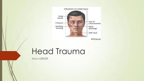
Head Trauma
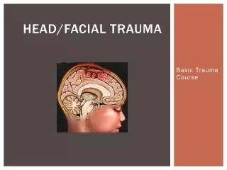
Head Trauma. Chapter 6. Objectives. Review the key anatomic features of the head (face) and neck Describe injury patterns Describe the evaluation of the patient with suspected head (face)/neck injury Review epidemiology and pathology associated with traumatic brain injury (TBI)


Head Trauma
E N D
Presentation Transcript
Head Trauma Chapter 6
Objectives • Review the key anatomic features of the head (face) and neck • Describe injury patterns • Describe the evaluation of the patient with suspected head (face)/neck injury • Review epidemiology and pathology associated with traumatic brain injury (TBI) • Understand the initial evaluation and management of a patient with a TBI
ANATOMY • Soft Tissues • Includes parotid glands • Bones • Facial and cervical spine • Neck blood vessels • Carotid and vertebral arteries • Jugular and other veins • Trachea • Esophagus • Globes and surrounding ducts
ABCDE’s REMAIN BASIC! • Soft tissue or bony injuries may immediately threaten the airway • Uncontrolled bleeding can change “stable” to “unstable” very quickly • Standard maneuvers may be less successful in setting of fractures, etc. • Associated brain or spine injuries may cause airway loss as well • All blunt face/neck trauma must be considered at risk for C-spine injury • Neurologic injuries may worsen with time as well
TRACHEAL INJURIES • Securing the airway remains critical • Tracheal injuries may cause significant air leak • Pneumomediastinum • Pneumothorax, even tension pneumothorax • Surgical repair is required • If unavailable, manage with secure airway and chest tubes if necessary • Minimize airway pressure on ventilator
BLUNT TRACHEAL INJURY Pneumothorax and Pneumomediastinum Tracheal Injury
EPISTAXIS • May result in significant bleeding • Separated into anterior and posterior sources • Intubation for airway control prior to packing may be needed
EPISTAXIS Posterior Packing
EPISTAXIS Anterior Packing Epistat Balloon
ZONE 1 INJURY • Difficult to access from neck incision, may need sternotomy/thoracotomy • Initial management with angio/CT angio, bronchoscopy, esophagoscopy • Basically need to evaluate all vascular and aerodigestive structures potentially in harm’s way • As with most trauma, “stable or unstable” guides the initial management • Active bleeding, expanding hematomas, or hemodynamic instability need to be addressed first in the OR and then with staged work up if indicated
ZONE 2 INJURY • Only zone that is easily accessed from a neck incision • Still requires investigation of vascular and aerodigestive structures • In a STABLE patient, can be investigated with CT and endoscopy potentially • Again, unstable patients or those with active bleeding issues need to be addressed in the operating room!
ZONE 3 INJURY • Similar to Zone I, potentially difficult to access surgically and so angiography or CT needed, with possible endoscopy • These tend to be vascular injuries at the skull base that are very difficult to control surgically • Again, instability should prompt rapid damage control in OR, followed by additional work up if needed
VASCULAR INJURY COMPLICATIONS • Hemorrhage is the first concern • Stroke is the second concern (up to 25% of ICA injuries) • Revascularization may be required ICA/ECA Injury with Reconstruction
BLUNT CEREBROVASCULAR INJURY • More frequent that was believed in the past • Roughly 1-1.5% of blunt admissions • Workup with CT Angio or conventional angiography • Treatment based on grade • Low grade lesions no intervention or ASA • Higher grade lesions need anticoagulation or possibly stenting, with recent interest in aggressive antiplatelet agents • Complications related to increased stroke risk
FACIAL FRACTURES • Frequent injuries, but rarely have to be addressed immediately from a surgical standpoint • The primary question should be one of airway protection • The anatomic disruption or bleeding may cause loss of airway • The situation may deteriorate as swelling progresses in the upper airway • Remember that the globes may be injured by fractures and a good exam, including visual acuity, is mandatory
UPPER FACE FRACTURES • Clinical exam is very useful – pain, bruising, crepitance, movement • Malocclusion often occurs with mandible fractures • Check a cranial nerve exam!
MANDIBLE FRACTURES • Malocclusion a common hint on exam • 50% will break multiple places • Can be managed with soft diet/liquids and pain control in short term • Operative repair ultimately required Panorex
FACIAL FRACTURES • Open fractures may require broad spectrum antibiotic coverage • This isn’t agreed upon, but if a sinus is violated then initial coverage is reasonable • Remember that if enough trauma occurred to fracture bones, the nearby structures are also at risk • At least 20% of facial fractures will have a TBI • About 2% will have a C-spine fracture
OCULAR INJURIES • Evaluation requires a careful exam, including visual acuity • Open globes are as emergent as threatened limbs, and need antibiotic coverage like open fractures • Remember that open globes need an altitude restriction for MEDEVAC
OCULAR INJURIES • Layering of blood in the inferior anterior chamber • Usually managed with rest, elevation of HOB, and correction of clotting factors • 5% will require surgical evacuation Hyphema
OCULAR INJURIES • Minor injury • Resolves spontaneously, though may take weeks • Avoid anticoagulant or antiplatelet drugs • Lubricant eye drops as needed Subconjunctival hemorrhage
SUMMARY thus far • Airway control remains the primary concern • Control of hemorrhage may require packing, angiography, or operation • Facial fracture repair may be delayed if necessary once wounds are closed • Tracheal and esophageal injuries require more urgent repair • Globe injuries should be considered with facial fractures, and known injuries treated with the same urgency as threatened limbs
GLASGOW COMA SCALE (GCS) EYE OPENING (E) Spontaneous 4 To Speech 3 To Pain 2 None 1 BEST MOTOR Obeys Commands 6 RESPONSE (M) Localizes Pain 5 Normal Flexion (Withdraws) 4 Abnormal Flexion (Decorticate) 3 Extension (Decerebrate) 2 None 1 VERBAL RESPONSE (V) Oriented 5 Confused Conversation 4 Inappropriate Words 3 Incomprehensible Sounds 2 None 1
HEAD INJURY • Mild: GCS 14 – 15 • Moderate: GCS 9 – 13 • Severe: GCS 3 - 8
STATISTICS • #1 Cause of Traumatic Death • 500,000 CHI per year • Upon ER Arrival: • Dead: 14% • Mild Injury: 80% • Moderate – Severe Injury: 10% each • Overall Mortality • Mild: 0% • Moderate: 7 – 10% • Severe: 30%
DISABILITY • CHI: Disability within each group • Mild: 10% • Moderate: 50 – 67% • Severe: > 95% • Penetrating GSW to Head • Vegetative or Severely Disabled: 10% • Moderate Disability: 20% • Good Outcome: 20% • The Others: Dead
PATHOLOGY • Anatomy • Types of Injury
ANATOMY • Pia Mater • Arachnoid Layer • Dura Mater
SUBDURAL HEMATOMA • Most common traumatic lesion • In 20-40% of severely head-injured • Located in space between dura and arachnoid layers • Venous bleed • Concave Shape
EPIDURAL HEMATOMA • Located in space between dura and inner table • Arterial bleed • Classic presentation • Convex shape
CEREBRAL CONTUSION • Most common in subfrontal and temporal regions • Bruised tissue with violation of BBB
DIFFUSE AXONAL INJURY (DAI) • Axonal structural failure due to mechanical forces along axons • Culmination in physical separation of axon into proximal and distal segments • Number of axonal disruptions = Amount of deficit
MANAGEMENT • Initial Evaluation • Emergent Surgical Management • Initial Management
EVALUATION • First Priority: Airway, Breathing, Circulation • Disability • Pupillary Exam • Calculate GCS • Resuscitation • Standard Techniques • DO NOT postpone treating hypotension • Preferred Crystalloid: Hypertonic Saline or Isotonic Saline • Blood
EVALUATION • Secondary Survey • Assess for other injuries • Complete neurological assessment • Radiologic Evaluation • Hemodynamically stable • CT Scan of Head: Gold Standard
EMERGENT SURGICAL INTERVENTION • Significant mass effect • Displacement of > 5mm off midline • Penetrating injuries may need simple debridement
INITIAL MANAGEMENT • Significant reductions in mortality & morbidity by using intensive management protocols: • Early intubation • Rapid transportation to appropriate trauma care facility • Prompt resuscitation • Early CT scanning • Immediate evacuation of intracranial mass lesions • TCDB Mortality: 50% vs 36% (using these protocols)
INITIAL MANAGEMENT • Sedation • No studies have proven sedation to influence TBI outcome • Neuromuscular Blockade – Results in: • Longer ICU Course • Increased pneumonia rate • Trend towards increased sepsis • No improvement in outcome
INITIAL MANAGEMENT • Blood Pressure • Single episode of hypotension (SBP < 90 mm Hg) • Increased morbidity • Doubled mortality • Hypotension is an INDEPENDENT predictor of outcome
INITIAL MANAGEMENT • Cerebral Perfusion Pressure • CPP = MAP – ICP • Very low following TBI; may be near ischemic threshold • Critical threshold is 60 mm Hg • Significant decline in outcome for those with persistent CPP < 60 mm Hg • CPP < 50 mm Hg associated with: • Critical reductions in PbO2 • Increased morbidity and mortality
INITIAL MANAGEMENT • Oxygenation • Hypoxemia: PaO2 < 60 mm Hg • Results in: • Increased mortality: 14% vs 50% • Worse outcome • Mannitol • Studies support use of mannitol for ICP management
INITIAL MANAGEMENT • Hyperventilation • Introduction • Cerebral blood flow in first day after injury is half that of normal individuals • Severe hyperventilation results in cerebral ischemia • Hyperventilation is known to decrease ICP and lower CBF • Aggressive Hyperventilation (PaCO2 < 30 mm Hg) • Reduces CBF, reduces ICP, possible loss of autoregulation • Outcomes at 3 and 6 months are better without prophylactic hyperventilation
INITIAL MANAGEMENT • Hyperventilation (continued) • Guidance • STANDARD: In absence of increased ICP, chronic prolonged hyperventilation (pCO2 < 35 mm Hg) therapy should be avoided after severe TBI • Guideline: Use of prophylactic hyperventilation during first 24 hours after severe TBI should be avoided because it can compromise CBF during a time when CBF is already low
ICP MONITORING • At Risk Patients • ICP Data and Patient Management • Guidelines
AT RISK PATIENTS • Mild and Moderate Head Injury • Low risk for ICH • Less than 3% (Mild) and 10 – 20% (Moderate) will deteriorate into coma • Routing ICP monitoring is not recommended • Severe Head Injury • ICH Incidence • Abnormal CT: 53 – 63% • Normal CT: 10 – 15% • Normal CT + 2 of 3 adverse features: Similar to abnormal CT • Normal CT Strategy • Up to 1/3 may develop new pathology within a few days from injury • Follow-up scanning for those without ICP monitoring
ICP DATA & MANAGEMENT • All Therapies are Double-Edged Swords • Hyperventilation • Reduces ICP • Causes cerebral vasoconstriction and ischemia • Mannitol • Reduces ICP • Cumulative doses can exacerbate brain edema • Sedation, analgesia, and paralysis • Reduce ICP • Impossible to interpret clinical exam
GUIDELINES • Severe TBI (after resuscitation) and Abnormal CT • Severe TBI (after resuscitation) and Normal CT + 2 of 3: • Age over 40 years • Unilateral or bilateral posturing • SBP < 90 mm Hg • Not routinely in Mild or Moderate TBI • ICP management initiated at upper threshold of 20 – 25 mm Hg