Download
1 / 37
370 likes | 503 Vues
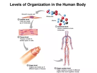
Organization of the Human Body. Chap 46. Cell specialization. Zygote Forms three germ layers Ectoderm; outher layer, skin & nervous system Mesoderm: middle layer, muscles, bones and connective tissue Endoderm: Inner layer, organs. Germ Layers. Cell specialization. Tissue types

E N D
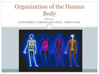
Organization of the Human Body Chap 46
Cell specialization • Zygote • Forms three germ layers • Ectoderm; outher layer, skin & nervous system • Mesoderm: middle layer, muscles, bones and connective tissue • Endoderm: Inner layer, organs
Cell specialization • Tissue types • Connective: binds and supports other structures (bones, elastins) • Function: gives strength to softer tissues • Origin: mesoderm
Cell specialization (cont) • Muscle • Function: movement • Origin: mesoderm
Cell specialization (cont) • Nerve • Function: communication, coordination, information • Origin: ectoderm • Types of nervous transmission • Afferent: sensory • Efferent: motor
Afferent Efferent Afferent vs Efferent nerves
Skeletal System • Endoskeleton • Advantages • Flexible • No molting • Greatest support with least weight • Disadvantages • less protection than exo- • Depend on nervous system for protection
Skeleton Functions • Support • Muscle attachment • Protection: organs and CNS • Store minerals • Marrow storage
Bone development • Long bones • Develop from cartilage: Ossification • Remove minerals from blood • Calcium phosphate ions • Calcium carbonate ions • Continues throughout childhood
Bone development (cont) • Flat bones: cranium, sternum, jaw • Develop from membrane layers • Sutures: joints in bones • Bones not fused in baby: allows brain to grow
Bone Structure • Diaphysis: shaft of bone • Epiphysis: ends of long bones • Epiphyseal line: growth line/plate • Haversian system • Haversian canal: channels through bones • Functions: delivers blood to bone, nutrients, osteocytes • Marrow • Red • Found: flat bones, epiphyseal portion of long bones • Function: RBC, WBC (erythrocytes, leucocytes) • Yellow • Found: central cavity of long bones (diaphysis) • Function: fat storage (in emergency may produce RBC’s
Joints • Types • Hinge: elbow • Ball and socket: hip • Angular: wrist • Gliding: vertebrae • Pivotal: Atlas and Axis • Top 2 vertebrae that allow head rotation • Attachment • Bone to bone: ligaments • Muscle to bone: tendons
Muscle systems • Muscle tissue • Made of cells that are able to shorten
Muscle fibers • Fibers are single cells • Energy provided by ATP (lots of mitochondria) • Bundle of fibers is a motor unit
Muscle contraction videos • http://www.youtube.com/watch?v=CepeYFvqmk4 • http://www.youtube.com/watch?v=NRzJjx3ANuE
Three Muscle types • (get notes from descriptions of pictures)
Flexors vs Extensors • Flexor- pulls bone toward each other (flex) • Extensors- pulls bones away from each other (extend) • Work in opposites- each flexor has and extensor to control the range of motion
Heart Contractions • Sinoatrial node (pacemaker): in right atrium, impulse start here and causes contraction • Atrioventricular node: Causes ventricle contraction • Atria contract, then ventricles, moves right to left
Electrocardiograms (EKG/ECG) Figure 8.15B, C