Download
1 / 33
330 likes | 355 Vues
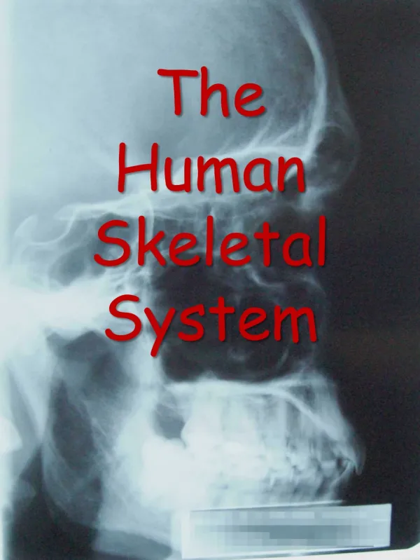
Skeletal System (View Bozeman Video). Quiz on labels. Quiz on labels. Handout 1 with vocabulary. Bone Development. Parts: Each end of a long bone is wider than the central portion of the bone. The enlarged end is called the Epiphysis

E N D
Parts: Each end of a long bone is wider than the central portion of the bone. The enlarged end is called the Epiphysis Between the epiphyses (ends of the bone), there is a narrow region of bone diaphysis (di af i sus). A covering of fibrous C.T. encloses all bone (except the areas covered by articular cartilage) and is called: Periosteum READ H.O. BONE Structure: Ex: Long Bone (First three paragraphs) See Handout on Bone Structure : Notes here for emphasis only
Bone Cell Types (see handout) Osteoprogenitor cells • Bone forming cells • Differentiate into osteoblasts
Osteoblasts/Osteocytes How do they differentiate? What changes? Matrix surrounds mature osteocytes
Osteoclasts • Function in bone reabsorption
Notes Begin here! Bone Development • Develops from mesoderm. • Begins 1st trimester • Formation of cartilage/ membrane skeleton occurs. • Membrane portions are actually undifferentiated connective tissue (CT). • End of third month: Large amount is replaced with bone • Bony structures continue to develop into adulthood.
Bones form in two ways (refer to diagram for emphasis, but take notes here) Intramembraneous ossificiation: • Called membrane bones • bone formed within undifferentiated CT. Ex. Bones of skull, mandible, clavicles Endochondral ossification • Called endochondral bones • This replaces existing cartilage model. Ex. Most bones of skeleton
Intramembranous ossification • Layers of undifferentiated C.T. form at site of future bones • Layers contain dense networks of blood vessels. • Osteoprogenitor cells form in two areas: • Around b.v. of C.T. • Along inner surface of C.T. • They then differentiate into osteoblasts This goes with NOTES Use this part of diagram for endochondral ossification to follow
Osteoblasts secrete bone matrix around themselves, forming trabeculae (cancellous bone) Trabeculae develop in all directions along b.v. and forms center of developing bone. Once osteoblasts form their own lacunae they become osteocytes. Canaliculiform and interconnect the osteocytes with each other and with osteoblasts along the surface. Use this part of diagram for endochondral ossification to follow
In addition… • Undifferentiated C.T. above/below developing cancellous bone form periosteum • Osteoblasts along inner surfaces of periosteum deposit matrix making layer of compact bone. • Osteoblasts also develop into osteocytes once they form their own lacunae.
Endochondral OssificationEX. USED = LONG BONE At beginning of first trimester, mesoderm differentiates into the cartilage “models” of the bone that is to be formed. Models are composed of hyaline cartilage and surrounded by perichondrium Why do you think we call these ‘models?’ Remember to see bottom of previous handout for further explanation…
By the third month, “ossification” process begins in areas of cartilage model called “ossification centers.” There are three ossification centers in a long bone, one "primary" and two "secondary". The primary ossification center (primary O.C.) is located in the center of the diaphysis. There is onesecondary ossification center (secondary O.C.) in each epiphysis. The replacement of the hyaline cartilage model by bonebegins in the primary O.C. and progresses in two directions toward the epiphyses. The secondary O.C. does not functionuntil later. These areas remain cartilage so that the skeleton can continue to grow along with the rest of the body.
PROCESS OF OSSIFICATION in LONG BONES • A ring of periosteum develops around the primary O.C. • Osteoblastsdevelop underneath this ring and produce a thin layer of compact bone that surrounds the primary O.C. • Some calciumbegins to move into the cartilage matrix located inside the ring, causing it to calcify. • When the cartilage matrix becomes calcified, the nutrient supply to the chondrocytes (cartilage cells) is cut off because diffusion cannot occur through a calcified matrix. • Then the chondrocytes die and disintegrate leaving open spaces in the calcified matrix. What happens to the open spaces???
Blood vessels, osteoprogenitor cells and undifferentiated C.T. cells move into the open spaces. • The blood vessels begin to branch and grow. • They eventually develop into the Haversian system. • The osteoprogenitor cells differentiate into osteoblasts and begin to secrete matrix forming cancellous bone. • The undifferentiated C.T. cells develop into marrow and later function in hematopoiesis. • The osteoblasts that develop inside (under) the periosteum continue to produce compact bone. Go to: Animation of Bone Ossification www.nursereview.org OR Utube: Process of ossification
This process of ossification continues in two directions towards the epiphyses. • This provides additional skeletal support to the developing fetus. • Cartilage formation also continues so that the overall size of the skeleton grows along with the rest of the body.
SECONDARY OSSIFICATION CENTERS SEE HANDOUT FOR THESE NOTES!!!
SEE HANDOUT Remaining areas are: 1. On the articulating surfaces of bone (This forms the articular cartilage that remains throughout life.) And.. 2. A region of cartilage remains in each end of the bone. These regions are located between the diaphysis and epiphyses. They are called epiphyseal disks / plates, or “growth plates”
The growth plates (epiphyseal disks) continue to produce cartilage while the individual grows postnatally. Each disk is divided into four layers
Continue with handouts from here to end…Read on your own starting with E and working forward (yellow packet)! • As long as the chondrocytes in the second layer undergo cell division, the bone grows in length. This occurs with the greatest speed between the ages of 5 – 12 years in females and 5 – 14 years in males, then it slows down. Eventually the primary and secondary ossification centers grow together because the chondrocytes in the second layer stop dividing. The growth plate becomes thinner and thinner until it is completely ossified and the bone can no longer grow in length. • By the ages of 17 – 20 years, the bones of the upper limbs are completely ossified. • By the ages of 18 – 23 years, the bones of the lower limbs are completely ossified. • Normally, by the age of 25 years all bone is completely ossified.
Bone diameter • The diameter of the bone must also increase as the bone grows in length. The diameter is increased when osteoblasts develop from the inside of the periosteum. They form compact bone which is added to the outer surface of the bone. • At the same time bone resorption occurs in the middle of the diaphysis. During this process, osteoclasts develop in the marrow cavity. These cells secrete enzymes that erode away the bone tissue in the center of the bone. This eliminates the trabeculae of the cancellous bone and hollows out the marrow cavity. This process (bone resorption) also releases calcium and other minerals for use by the body. Normally the cancellous bone in the epiphyses is not resorbed, because it is needed for strength.
Bone Remodeling / Bone Homeostasis "Bone remodeling" is the ongoing replacement of old bone tissue by new bone tissue. This occurs throughout life and occurs at different rates in different bones. EX: The distal end of the femur is replaced about every 4 months, yet the middle of the diaphyses of some long bones are never fully replaced. This process redistributes the bone matrix along lines of mechanical stress. It is the result of the alternate actions of osteoclasts (bone resorption) and osteoblasts (bone replacement).
Read on Your own: Factors Affecting Bone Growth and Development and Blood Cell Formation(handout)Fractures/Osteoporosis reading and questions follow! See www.nursereview.org ORsearch “Fracture repair”
Continue with R.S. 9 Bone packet to follow on the Human Skeleton and will reflect R.S. 10