Download
1 / 34
430 likes | 1.98k Vues
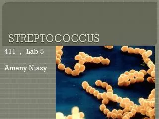
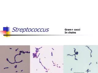
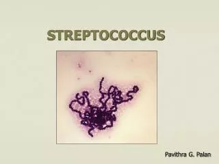
Practical No.10 Streptococcus species. GENERAL CHARACTERISTIC:. * G+ve cocci , arrange in chains or pairs. * Some strains are capsulated * Majority are facultative anaerobic, few are obligatory anaerobic. * Catalase –ve * Non motile. * Non spore forming * Fastidious microorganism.

E N D
Practical No.10 Streptococcus species
GENERAL CHARACTERISTIC: * G+ve cocci , arrange in chains or pairs. * Some strains are capsulated * Majority are facultative anaerobic, few are obligatory anaerobic. * Catalase –ve * Non motile. * Non spore forming * Fastidious microorganism
Lab dx. Specimens: sputum, throat swab, nasopharyngeal swab, blood, CSF…etc. Gram stain: G+ve cocci, arrange in chains. Culture: on blood agarpinpointed, Grayish white, translucent, matte or glossy colonies with large zone of β- hemolysis. Bacitracin disc (0.04 U) sensitivecauses zone of growth inhibition. Serology: Lancefield grouping, M-protein serotyping and ASO test (Antistreptolysin-O test).
Members of the genus Streptococcus are responsible for disease as well as being part of the normal flora of humans. Among the diseases caused are: • bacterial pneumonia • meningitis • Tonsillitis • Endocarditis • scarlet fever • Erysipelas • urinary tract infections. • Streptococcus species are also found normally in the mouth and on the skin surface. • The streptococci are classified by two major methods: • hemolytic activity • serologic classification of Lancefield.
When grown on sheep blood agar, streptococci display one of three types of hemolysis of the red blood cells in the agar. • Alpha hemolysis--The red blood cells in the media are partially digested producing a greening of the agar. • Beta hemolysis--The red blood cells in the media are completely digested producing a clearing of the agar. • Gamma hemolysis--No change is noted in the agar. The red blood cells are not lysed. • Expected Hemolysis: • Streptococcus pyogenes always beta hemolylic • Streptococcus agalactiaeusually beta hemolytic • Streptococcus pneumoniaeandViridans streptococciare always alpha hemolylic • Enterococcus faecalisgamma hemolytic
(Beta hemolysis) clear zone around colonies S.pyogenes on blood agar (beta hemolysis)
Classification Based on Lancefield Proteins: Rebecca Lancefield, working with various streptococcal species, discovered proteins in the cell wall that were unique to certain organisms. These proteins were labeled Group A, Group B, Group C, and so on through Group M. Currently three Lancefield Groups are of medical importance: Group A, Group B, and Group D Group A Strep--Streptococcus pyogenes Group B Strep--Streptococcus agalactiae Group D Strep--Streptococcus bovis, Enterococcus (Streptococcus) faecalis Streptococcus pneumoniaedoes not possess Lancefield proteins and is not classified in one of the Lancefield groups. Viridans streptococci are the term applied to alpha hemolytic Streptococcus species that lack Lancefield proteins.
Beta hemolytic streptococci: Streptococcus pyogenes (Group A strep) are gram-positive cocci usually in chains.The group A streptococcus bacterium (Streptococcus pyogenes, or GABHS) is a form of β-hemolytic Streptococcus bacteria responsible for most cases of streptococcal illness. Other types (B, C, D, and G) may also cause infection such as: bacteremia , impetigo, cellulitis, erysipelasosteomyelitis, sinusitis, tonsillitisseptic arthritis ,pneumonia, necrotizing fasciitis , meningitis, scarlet fever strep throattoxic shock syndrome. Streptococcus pyogenes
S.pyogenes (Group A β- hemolytic,GABH): M-protein 80 serotypes Transmission Spread by respiratory droplets or direct contact Reservoir Human throat and skin(N.F.)
Identification of group A Streptococci. (Streptococcus pyogenes). Specimen; pus, blood, serum, throat swab, nasal swab. Bacitracin test: Commercially available paper disks saturated with a solution containing Bacitracin will inhibit about 97% of all strains of Group A streptococci; other groups of beta-hemolytic streptococci will not be affected. Procedure; Streak a blood agar plate with an isolated colony of beta-hemolytic streptococci. After inoculation, flame the provided forceps, and aseptically pick up a bacitracin disk (B or A disk). Place the disk on the plate and press gently onto the agar medium to ensure firm contact with the agar. Observe the plates for inhibition of growth (indicating sensitivity) after overnight incubation at 37. For Streptococcus pyogenes there will be a zone of inhibition of growth around the A disk.
Phadebact Strep (Coagglutination) test: The Phadebact brand rapid streptococcal identification system is based on a coagglutination reaction. A patient sample or bacteria from a culture are mixed with a solution that contains antibody to the Group A antigenic determinant of the S. pyogenescell. The antibodies are bound through their Fc portion to nonviable Staph aureuscells via protein A, so that if the antibodies bind at their Fab site to group A streptococci, the staphylococci will be clumped together as a lattice of immune complexes forms (coagglutination) If no Group A streptococci are present, the staphylococci will remain in a homogeneous suspension. Note that this test is looking for antigen from the patient sample, not antibodies. The antibodies are a reagent of the test kit.
Procedure: • a. Mix the two reagent solutions by vigorous shaking to suspend the staphylococcal cells. • b. Add one drop of the test reagent (A) to the oval for the test and one drop of the negative control reagent (-) to the oval beneath it. Make one set of test and contol drops for each unknown bacterium. • c. Using a sterile inoculating loop, pick about 5 colonies of the beta hemolytic streptococci and smear the cells into the two oval areas with the A and - reagent. • d. Stir the cells in the reagent, but be sure to use a new loop between the test and control samples to prevent cross-mixing of the reagents. Rock the card to continue mixing the samples. • e. If the test sample agglutinates, but the control sample does not, the sample is Group A streptococcus (Streptococcus pyogenes). If the test and unknown samples do not agglutinate, the test sample is not Group A streptococcus. In this figure the A reagent is across the top and the - reagent is across the bottom. The left two oval have a negative strain (non-Group A streptococcus) while the right two oval have Group A streptococcus -Streptococcus pyogene
The left 2 samples are a non-Group A strain, Note the agglutination reaction in the upper right and the lack of agglutination in the lower right.
AntistreptolysinO titre: ASO titre - titre of (serum) antistreptolysin O antibodies is a blood test used to assist in the diagnosis of a streptococcal infection or indicate a past exposure to streptococci.The ASOT helps direct the antimicrobial treatment and is used to assist in the diagnosis of scarlet fever, rheumatic fever and post infectious glomerulonephritis. A positive test usually is >200 units/mL, but normal ranges vary from laboratory to laboratory and by age. Normal levelsis less than 160 unit per milliliter,
ASO test: * Measure Ab against Streptolysin O *ASO test uses in post streptococcal infection complication. This test used to determine significance streptococcal infection by measuring the ASOT: * ASOT (Ab Titer): Normal < 200 < significance result
Identification of group B streptococci. (S.agalactiae) The CAMP Test The laboratory identification of Group B hemolytic streptococci has been simplified by implementation of the CAMP test which is easy to perform and well within the capabilities of small laboratories. The hemolytic activity of staphylococcal-hemolysin on RBC is enhanced by an extracellular factor produced by Group B streptococci, called the CAMP factor. Therefore, wherever the two reactants overlap in sheep BAP, an accentuation of the ß-hemolytic reaction will be noted.
Procedure: The CAMP test is performed by making a single streak of the streptococcus perpendicular to a strain of Staphylococcus aureus that is known to produce ß-hemolysin. The two streak lines must not touch one another. The inoculated plate must be incubated with an ambient atmosphere (room temperature). The plate should not be incubated in an anaerobic environment because many Group A streptococci are positive in the absence of O2. Any Bacitracin-negative, CAMP-positive, bile-esculin-negative streptococcus can be reported as: Group B streptococcus, presumptive by CAMP. Positive control: S. agalactiae.
The Hippurate test: is a qualitative procedure for determining the ability of bacteria to enzymatically hydrolyze sodium hippurate.;It is used in conjunction with other biochemical tests to identify cultures of of group B streptococci.
Procedure: 1. To a Hippurate Test tube, add 0.2ml (3-4 drops) of distilled water at a pH of 6.8-7.2. 2. Using a heavy inoculum (a full 10ul loop is adequate) from an 18-24 hour culture, make a heavy suspension of the organism in the Hippurate Reagent with a standard inoculating loop. 3. Incubate the tube for two hours at 35-37degrees C. 4. During the incubation period, reconstitute the Ninhydrin Indicator Solution in the dropper bottle by adding 2ml of distilled water at a pH of 6.8-7.2. Replace the cap tip and cap, and vigorously shake for one minute. Let stand at room temperature for 30 minutes or until all the substrate has dissolved. 5. After the two hour incubation period, add two drops of the Ninhydrin Indicator Solution to the Hippurate Reagent and organism mixture. 6. Reincubate at 35-37degrees C. for 30 minutes. Observe the tubes at 10 minute intervals for the appearance of a deep blue color, which is a positive test. The color change will usually appear in 10 to 15 minutes after the Ninhydrin Indicator Solution has been added.
Non-beta hemolytic (alpha- and nonhemolytic) streptococci The non-beta hemolytic streptococci include the S. pneumoniae, viridans streptococci, Group D streptococci and enterococci. Identification of group D Streptococci: Lancefield Group D streptococci are divided into two groups: (1) Enterococci, and (2) nonenterococci. Enterococcus faecalis and Enterococcus faecium are the species of Enterococcus . Strep. bovis and Strep. equinus are species of "Group-D streptococci, not Enterococci".
Bile-Esculin Hydrolysis Test: The purpose of this test is to determine the ability of an organism to hydrolyze the glycoside esculin to esculatin and glucose in the presence of bile (10 - 40%). This test aids in the differentiation of group D streptococci from other "not group D streptococci". Procedure: 1. Inoculate the organism to be tested into the bile esculin medium. Incubate at 37oC for 24 hours (stab into medium,then streak on slant). Positive Test: Presence of a black to dark brown color on the slant -(Enterococcusfaecalis) Negative Test: No blackening of the medium - (Streptococcusagalactiae or Streptococcus pyogenes)
Salt Tolerance Test for Enterococci This test is based on the ability of the enterococci to grow in 6.5% NaCl and separates them from the "Group-D streptococci, not enterococci". Procedure: 1. Label a high salt broth tube with the organism to be tested and your initials. 2. Using a sterile loop transfer the organism to be tested to the broth. 3. Incubate the tube for a minimum of 18 hours. 4. Examine the tube for evidence of growth (turbidity). Compare the tube to an uninoculated tube. Organisms that can tolerate a high salt environment (6.5% NaCl) will grow in this broth causing the broth to become cloudy or turbid. Turbidity is considered positive (+) for this test. Organisms that can not tolerate the high salt environment will not grow and the broth will remain clear. Clear broth is considered negative (-) for this test. Positive control: Enterococcus faecalis.