Download
1 / 31
330 likes | 464 Vues
Learn about the causes and types of cardiac arrhythmias such as heart blocks, sinus rhythm abnormalities, ectopic beats, and tachyarrhythmias. Explore ECG changes, symptoms, and treatment options.

E N D
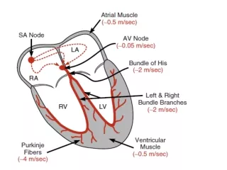
Causes of cardiac arrhythmias • Abnormal rhythmicity of the pacemaker • Shift of the pacemaker from the sinus node to another place of the heart • Blocks of different points in the spread of the impulse through the heart • Abnormal pathways of impulse transmission through the heart • Spontaneous generation of spurious impulses in almost any part of the heart.
Heart block: I. Block at the level of AV node: A. First degree heart block: every atrial depolarization is followed by conduction to ventricle but delay. ECG changes prolongation of PR interval to more than 0.22 second.
B. Second degree heart block: some P waves conducted but other not. ECG changes every second or third P wave conducted to the ventricles.
C. Third degree heart block (complete heart block): Rate:Atrial: 60–100 bpm; ventricular: 40–60 bpm Rhythm: Usually regular, but atria and ventricles act independently. It occurs when all atrial activity fails to conduct to the ventricle so the Bundle of His will be responsible form generation of impulses.
Caused by: Acute myocardial infarction, calcify aortic stenosis, cardiomyopathy, drugs (digoxin). Block below AV node: A. block at Bundle of His, B. Block at the branches (Right or Left branch).
Sinus rhythm: It is caused by the changes of number of impulses emitted form SA node. Heart rates more than 100/min is called (tachycardia), while less than 60/min is called (bradycardia). It is usually of two types: 1. Sinus bradycardia: Causes: A. Extrinsic causes: hypothermia, hypothyroidism, and raised intra cranial pressure, drugs (beta-blockers, digitalis, and anti-arrhythmic drugs). B. Intrinsic causes: acute ischemia, infarction of SA node. ECG changes: Prolonged R-R interval.
2. Sinus tachycardia: causes A. acute causes: exercise, emotion, pain, fever, acute heart failure, B. chronic causes: pregnancy, anemia, hyperthyroidism, excess catecholamine. ECG: short R-R interval.
Ectopic beat (extra-systoles, premature beat): A premature contraction is contraction of heart before the time that normal contraction would have been expected. Most premature contraction result from ectopic foci in the heart, which emits abnormal impulses at odd time during cardiac rhythm. Possible causes of ectopic foci are: Local area of ischemia Small calcified plaques at different points in the heart, which press against the adjacent cardiac muscle so some fibers are irritated Toxic irritation of the AV node, Purkinje system, or myocardium caused by drugs, nicotine, or caffeine. If an irritable ectopic focus discharges once, the result is ectopic beat. If the ectopic foci discharge repetitively at rate higher than that of SA node, it produces rapid, irregular tachycardia.
It could be: 1. Atrial ectopic: The ECG changes are: The P wave of this beat occurs too soon in the heart cycle, The P-R interval is shortened, indicating that the ectopic origin of the beat is near the A-V node The interval between the premature and the next succeeding contraction is slightly prolonged, which is called (compensatory pause).
2. ventricular ectopic: ECG changes: Premature beats that originate in an ectopic ventricular focus usually have bizarrely shaped prolonged and high voltage QRS complex The P wave is usually buried in the QRS of the extra-systole The T wave has an electrical potential polarity opposite to the QRS
Tachy-arrhythmia: Cardiac arrhythmia is a disturbance in electrical rhythm of the heart; this may be paroxysmal or continuous, and may cause sudden death, syncope, heart failure, palpitation, or no symptoms. There are two mechanisms for tachycardia: 1. Increase automaticity (increase slop angle): when the tachycardia is sustained by repeated spontaneous de-polarization of an ectopic focus or single cell.
2. Re-entry: when the tachycardia is initiated by an ectopic beat but sustained by a closed loop or re-entry circuit. Most tachy-arrhythmias are due to re-entry.
Causes of re –entry(circus movement) • Long pathway around the circle • Decreased velocity of conduction • Shortened refractory period of the muscle
The types of tachy-arhythmias are: I. Atrial tachy-arrhythmias: Causes: ischemic heart disease, Mitral valve disease, rheumatic heart disease, hypertension, cardio-myo-pathy, thyro-toxicosis, atrial septal defect, acute and chronic alcohol abuse pulmonary embolus.
A. Atrial fibrillation: ECG: normal but irregular QRS, there are no P waves but base line may show irregular fibrillation waves.
B. Atrial flutter: ECG: regular saw-tooth-like atrial flutters waves (F waves) between QRST complexes; with rate about 300 beat/min. the QRS conducted 150 if every other one is conducted.
C. Atrial tachy-cardia: An ectopic arial tachycardia due to increase automaticity is rare but is sometimes is manifestation of digitalis toxicity. Rate: 150–250 bpm, Rhythm: Regular P Waves: Normal (upright and uniform) but differ in shape from sinus P waves
III. Ventricular tachy-arrhythmia: A. Ventricular tachycardia: it is usually a serious condition because: This type of tachycardia dose not occurs unless considerable ischemic damage is present in the ventricles Ventricular tachycardia frequently initiates the lethal condition of ventricular fibrillation Cardiac output is decreased. The ECG changes including: a series of ventricular premature beats occurring one after another without any normal beat interspersed so QRS morphology is regular, the rate is between (140-220/min).
B. Ventricular fibrillation: The effects of ventricular fibrillation: The fibrillating ventricles, like the fibrillating atria, look like a quivering "bag of worms". The fibrillating ventricles cannot pump blood effectively and circulation of the blood stops. Therefore, in the absence of emergency treatment, ventricular fibrillation that last more than a few minutes is fatal. The most common cause of sudden death in patients with myocardial infarction is ventricular fibrillation. The ventricular fibrillation can often be stopped and converted to normal sinus rhythm by mean of electrical shock. The ECG changes: it shows undulating waves of varying frequency and amplitude.
Anti-arrhythmic drugs: Classification of anti-arrhythmic drugs (Vaughan-Williams classification): Goal of therapy: a. Therapy aimsto restore normal pacemaker activity modify impaired conduction that leads to arrhythmias. Conduction velocity depens on the size of the inward current during upstroke of the action potential (↑inward currernt→↑. The evocity of conductance)
b. Therapeutic effects are achieved by: sodium- or calcium-channel blockade, prolongation of effective refractory period (it is slightly longer than absolute refratory period), blockade of sympathetic effects on the heart. Many anti-arrhythmic drugs affect depolarized tissue (ectopic foci) to a greater extent than they affect normally polarized tissue.
Anti-arrhythmic Drugs Treatment of tachy-arrhythmias: Class I a. Quinidine b. Disopyramide c. Lidocaine [Xylocaine] d. Flecainide e. propafenone
Class II Mechanism: Class II drugs are β-adrenoceptor antagonists, including propranolol, which act by reducing sympathetic stimulation. They inhibit phase 4 depolarization, depress automaticity; prolong AV conduction, and decrease heart rate (except for agents that have sympathomimetic activity) and contractility. Major drugs: a. Propranolol [Inderal], b. atenolol, c.Metoprolol d. Bisoprolol e. sotalol
Class III Class III drugs prolong action potential duration and effective refractory period. These drugs act by interfering with outward K currents or slow inward Na currents 1. Amiodarone [Cordarone]: a. Amiodarone is structurally related to thyroxine. It increases refractoriness, and it also depresses sinus node automaticity and slows conduction.
Class IV drugs Mechanism a. Class IV drugs selectively block L-type calcium channels. b. These drugs prolong nodal conduction and effective refractory period and have predominate actions in nodal tissues Verapamil [Calan,Isoptin]: a. Verapamil is a phenylalkylamine that blocks both activated and inactivated slow calcium channels.
Other anti-arrhythimic drugs • Digoxin: can control ventricular response in atrial flutter or fibrillation. • Digoxin toxicity • Extracardiac manifestations • a. anorexia, nausea, vomiting • b. Diarrhoea • cardiac manifestations • Bradycardia • b. Multiple ventriclarectopics • c. Ventricular bigeminy
Treatment of Brady-arrhythmia: 1. Atropine a. Atropine blocks the effects of acetylcholine. It elevates sinus rate and AV nodal and sinoatrial (SA) conduction velocity, and it decreases refractory period. b. Atropine is used to treat bradyarrhythmias that accompany MI. 2. Isoproterenol [Isuprel] a. Isoproterenol stimulates β-adrenoceptors and increases heart rate and contractility. b. Isoproterenol is used to maintain adequate heart rate and cardiac output in patients with AV block.