Download
1 / 63
640 likes | 700 Vues
Learn to identify, discuss etiologies, and manage common cardiac arrhythmias - sinus bradycardia, tachycardia, Arrhythmia, Sick Sinus Syndrome, PSVT, AFib. Treatment guidelines included.

E N D
Cardiac Arrhythmias Elise Georgi Morris, M.D. June 5, 2007
Objectives • Identify common arrhythmias encountered by the family physician • Discuss arrhythmia etiologies • Discuss initial primary care work-up and treatment • Practice questions
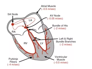
Normal Sinus Rhythm www.uptodate.com Implies normal sequence of conduction, originating in the sinus node and proceeding to the ventricles via the AV node and His-Purkinje system. EKG Characteristics: Regular narrow-complex rhythm Rate 60-100 bpm Each QRS complex is proceeded by a P wave P wave is upright in lead II & downgoing in lead aVR
Sinus Bradycardia • HR< 60 bpm; every QRS narrow, preceded by p wave • Can be normal in well-conditioned athletes • HR can be<30 bpm in children, young adults during sleep, with up to 2 sec pauses
Sinus bradycardia--etiologies • Normal aging • 15-25% Acute MI, esp. affecting inferior wall • Hypothyroidism, infiltrative diseases (sarcoid, amyloid) • Hypothermia, hypokalemia • SLE, collagen vasc diseases • Situational: micturation, coughing • Drugs: beta-blockers, digitalis, calcium channel blockers, amiodarone, cimetidine, lithium
Sinus bradycardia--treatment • No treatment if asymptomatic • Sxs include chest pain (from coronary hypoperfusion), syncope, dizziness • Office: Evaluate medicine regimen—stop all drugs that may cause • Bradycardia associated with MI will often resolve as MI is resolving; will not be the sole sxs of MI • ER: Atropine if hemodynamic compromise, syncope, chest pain • Pacing
Sinus tachycardia • HR > 100 bpm, regular • Often difficult to distinguish p and t waves
Fever Hyperthyroidism Effective volume depletion Anxiety Pheochromocytoma Sepsis Anemia Exposure to stimulants (nicotine, caffeine) or illicit drugs Hypotension and shock Pulmonary embolism Acute coronary ischemia and myocardial infarction Heart failure Chronic pulmonary disease Hypoxia Sinus tachycardia--etiologies
Sinus Tachycardia--treatment • Office: evaluate/treat potential etiology :check TSH, CBC, optimize CHF or COPD regimen, evaluate recent OTC drugs • Verify it is sinus rhythm • If no etiology is found and is bothersome to patients, can treat with beta-blocker
Sinus Arrhythmia • Variations in the cycle lengths between p waves/ QRS complexes • Will often sound irregular on exam • Normal p waves, PR interval, normal, narrow QRS
Sinus arrhythmia • Usually respiratory--Increase in heart rate during inspiration • Exaggerated in children, young adults and athletes—decreases with age • Usually asymptomatic, no treatment or referral • Can be non-respiratory, often in normal or diseased heart, seen in digitalis toxicity • Referral may be necessary if not clearly respiratory, history of heart disease
Sick Sinus Syndrome • All result in bradycardia • Sinus bradycardia (rate of ~43 bpm) with a sinus pause • Often result of tachy-brady syndrome: where a burst of atrial tachycardia (such as afib) is then followed by a long, symptomatic sinus pause/arrest, with no breakthrough junctional rhythm.
Sick Sinus Syndrome--etiology • Often due to sinus node fibrosis, SNode arterial atherosclerosis, inflammation (Rheumatic fever, amyloid, sarcoid) • Occurs in congenital and acquired heart disease and after surgery • Hypothyroidism, hypothermia • Drugs: digitalis, lithium, cimetidine, methyldopa, reserpine, clonidine, amiodarone • Most patients are elderly, may or may not have symptoms
Sick sinus syndrome--treatment • Address and treat cardiac conditions • Review med list, TSH • Pacemaker for most is required
Paroxysmal Supraventricular Tachycardia • Refers to supraventricular tachycardia other than afib, aflutter and MAT • Occurs in 35 per 100,000 person-years • Usually due to reentry—AVNRT or AVRT
PSVT • Initial eval: Is the patient stable? • Determine quickly if sinus rhythm • If not sinus and unstable, cardioversion • Unstable sinus tachycardia---IV beta-blocker, and treat cause • Sxs of instability would include: chest pain, decreased consciousness, short of breath, shock, hypotension—unstable sxs require shock
PSVT • If stable, determine whether regular rhythm (sinus or PSVT) vs irregular (afib/flutter, MAT)? p waves (MAT vs. AF)? • If regular, determine whether p waves are present, if can’t see---administer adenosine (6mg, can give 2 doses) or CSM or other vagal maneuvers)
PSVT • CSM or adenosine commonly terminate the arrhythmia, esp, AVRT or AVNRT • Can also use CCB or beta blockers to terminate, if available • Counsel to avoid triggers, caffeine, Etoh, pseudoephedrine, stress
PSVT • No p waves —junctional tachycardia, AVRT or AVNRT, Afib • AVRT and AVNRT: can have retrograde p waves and short RP interval • Abnormal p waves morphology: MAT
Atrial Fibrillation • Irregular rhythm • Absence of definite p waves • Narrow QRS • Can be accompanied by rapid ventricular response
Hypertension Hyperthyroidism and subclinical hyperthyroidism CHF (10-30%), CAD Uncommon presentation of ACS Mitral and tricuspid valve disease Hypertrophic cardiomyopathy COPD OSA ETOH Caffeine Digitalis Familial Congenital (ASD) Atrial Fibrillation—causes and associations
Atrial fibrillation--assessment • H & P—assess heart rate, sxs of SOB, chest pain, edema (signs of failure) • If unstable, need to cardiovert • Echocardiogram to evaluate valvular and overall function • Check TSH • Assess for RVR • Assess onset of sxs—in the last 24-48 hours? Sudden onset? Or no sxs?
Atrial fibrillation--management • Rhythm vs Rate control—if onset is within last 24-48 hours, may be able to arrange cardioversion—use heparin around procedure • Need TEE if valvular disease (high risk of thrombus) • If unable to definitely conclude onset in last 24-48 hours: need 4-6 weeks of anticoagulation prior to cardioversion, and warfarin for 4-12 weeks after
Atrial Fibrillation • Cardioversion: synchronized (w/QRS) delivery of current to heart; depolarizes tissue in a reentrant circuit; afib involves more cardiac tissue, but cardiovert • Defibrillation: non-synchronized delivery of current
Atrial fibrillation--management • Rate control with chronic anticoagulation is recommended for first line approach for majority of patients; overall Afib is a stable rhythm • Beta-blockers (atenolol and metoprolol) or calcium channel blockers (verapamil or diltiazem) recommended. Digoxin not recommended for rate control • Anticoagulation: LMWH and then warfarin; can use aspirin for anticoagulation if CI to warfarin, not as effective
Atrial fibrillation--management • Goal INR of 2.5 (2.0-3.0) • Rhythm control---second line approach, if unable to control rate or pt with persistent sxs • Can also consider radiofrequency ablation at pulm veins
PAC • P wave from another atrial focus • Occurs earlier in cycle • Different morphology of p wave
PAC • Benign, common cause of perceived irregular rhythm • Can cause sxs: “skipping” beats, palpitations • No treatment, reassurance • With sxs, may advise to stop smoking, decrease caffeine and ETOH • Can use beta-blockers to reduce frequency
1st Degree AV Block • PR interval >200ms • If accompanied by wide QRS, refer to cardiology, high risk of progression to 2nd and 3rd deg block • Otherwise, benign if asymptomatic
2nd Degree AV Block Mobitz type I (Wenckebach) • Progressive PR longation, with eventual non-conduction of a p wave • May be in 2:1 or 3:1
Wenckebach, Mobitz type I • Usually asymptomatic, but with accompanying bradycardia can cause angina, syncope esp in elderly—will need pacing if sxs • Also can be caused by drugs that slow conduction (BB, CCB, dig) • 2-10% long distance runners • Correct if reversible cause, avoid meds that block conduction
2nd degree block Type II (Mobitz 2) • Normal PR intervals with sudden failure of a p wave to conduct • Usually below AV node and accompanied by BBB or fascicular block • Often causes pre/syncope; exercise worsens sxs • Generally need pacing, possibly urgently if symptomatic
3rd Degree AV Block • Complete AV disassociation, HR is a ventricular rate • Will often cause dizziness, syncope, angina, heart failure • Can degenerate to Vtach and Vfib • Will need pacing, urgent referral
PVC • Extremely common throughout the population, both with and without heart disease • Usually asymptomatic, except rarely dizziness or fatigue in patients that have frequent PVCs and significant LV dysfunction
PVC • No treatment is necessary, risk outweighs benefit • Reassurance • Optimize cardiac and pulmonary disease management
Non-sustained Ventricular tachycardia • Defined as 3 or more consecutive ventricular beats • Rate of >120 bpm, lasting less than 30 seconds • May be discovered on Holter, or other exercise testing
Non-sustained ventricular tachycardia • Need to exclude heart disease with Echo and stress testing • If normal, there is no increased risk of death • May need anti-arrhythmia treatment if sxs • In presence of heart disease, increased risk of sudden death • Need referral for EPS and/or prolonged Holter monitoring
Ventricular fibrillation • Defibrillation
Practice Questions—Case 1 • 37-year old male comes to office for “skipping heart beats.” Going on over last 8 months, no other sxs: no sweating, palpitations, wt loss, chest pain, anxiety or pleuritic chest pain. On PE, BP is 100/70, normal S1S2, no murmurs/gallops. You hear about 5 premature beats.
Practice Questions • What is the most commonly encountered “premature contraction?” a. a ventricular premature beat b. an atrial premature beat c. atrial flutter d. atrial fibrillation e. none of the above
Practice questions Answer B: atrial premature beats • Most common premature beat in adults • Almost always asxs • Often patients c/of sxs during stress, or while laying quietly
Practice Questions 2. Most atrial premature beats discovered on clinical examination are: a. associated with COPD b. completely benign c. associated with valvular heart disease d. associated with an increase in cardiovascular mortality e. None of the above
Practice Questions Answer B: completely benign • require no treatment except reassurance
Practice Questions 3. Most ventricular premature beats discovered on clinical exam are: a. associated with COPD b. completely benign c. associated with valvular heart disease d. associated with increased cardiovascular mortality e. none of the above
Practice Questions Answer B: completely benign • Patients need reassurance • Usually asymptomatic • Occasionally can be associated with severe heart disease with multiple PVCs in a row---Vtach---which can cause syncope, chest pain, dyspnea and cardiac arrest
Practice Questions—Case 2 A 51 year old male presents to the emergency room with an acute episode of chest pain. He has a history of afib. On exam BP is 70/50, and ventricular rate is 160. He is in acute distress. His resp rate is 32. ECG shows afib with rapid ventricular response.
Practice Questions 4. What should your first step in management be? a. digitalize the patient b. give the patient IV verapamil c. give the patient IV adenosine d. start synchronized cardioversion e. start rapid IV hydration
Practice Questions Answer D: this patient has an acute onset afib with RVR, with chest pain and hypotension. Treatment of choice per ACLS protocol is cardioversion.
Practice Questions—Case 3 A 44 year old male comes to your ER saying he has palpitations. Denies chest pain or SOB. No known history of CAD or risk factors except mild obesity. Does admit to drinking heavily the night before. On PE, BP is 120/80 and heart rate is 160. ECG confirms afib with rapid ventricular response.
Practice Questions 5. What should you do at this time? a. digitalize the patient b. treat the patient with IV verapamil c. treat the patient with IV procainamide d. cardiovert the patient e. have him perform a Valsalva maneuver by rebreathing into a paper bag