Download
1 / 59
640 likes | 949 Vues
Cardiac Arrhythmias. A Guide For Medical Students William Beaumont Hospital Department of Emergency Medicine. In evaluating arrhythmias -. Rate - Is it fast or is it slow If slow – is there group to group beating Rhythm - Is it regular, irregular or irregularly irregular?

E N D
Cardiac Arrhythmias A Guide For Medical Students William Beaumont Hospital Department of Emergency Medicine
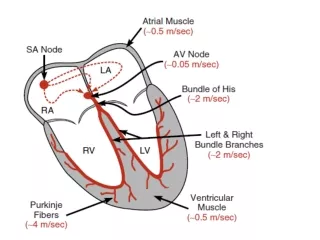
In evaluating arrhythmias - • Rate - Is it fast or is it slow • If slow – is there group to group beating • Rhythm - Is it regular, irregular or irregularly irregular? • P waves - Are they present? • QRS - Is it narrow or wide?
Sinus Bradycardia • What is it? • What causes it? • When do you treat it? • How do you treat it?
Sinus Bradycardia • A sinus rhythm with normal intervals and a rate less than 60 bpm • Normal variant, beta blocker overdose, dig, hypothermia, hypothyroidism, brady-tachy syndrome, and SA node ischemia • Requires treatment only if there is evidence of hypoperfusion • Two treatment options • Pacing: transvenous or transcutaneous • Atropine 0.5 mg IVP
Sinus Tachycardia • A sinus rhythm faster than 100 bpm • Etiology - usually a physiologic response to a stressor • Volume depletion / low stroke volume • Hypoxia • Systemic pathology: fever, anemia, hyperthyroidism • Drugs • Treatment - treat the underlying cause
Atrial Arrhythmias PACs MAT Atrial Fibrillation, atrial flutter SVT
Multifocal Atrial Tachycardia • Diagnosis requires the presence of three distinct p waves in a narrow complex tachycardia • Almost always associated with pulmonary disease • Less often due to hypokalemia or hypomagnesemia • Treat the underlying disorder – usually hypoxia • Unlike the other atrial tachyarrhythmias, cardioversion is of no value in MAT
MAT Rule of Threes 3different p waves, 3different pr intervals and 3 different r to r intervals
Causes of A-fib • Cardiovascular - CAD, HTN, CHF, myopathy, myo-, endo- and pericarditis, infiltrative disease, valvular, congenital • Metabolic - thyroid, electrolytes • Pulmonary - pulmonary HTN, PE • Toxic - cocaine, ETOH (holiday heart), beta agonists • Sepsis • Idiopathic
ECG Rules for A-fib • Regularity - irregularly irregular • Rate - atrial rate usually > 350 • Controlled - ventricular rate < 100 • RVR - ventricular rate > 100 • P wave - none discernable, may be f waves • QRS - less that 0.12 seconds (easy dx). If > 0.12 sec must rule out VT (which is usually more regular)
A-fib with RVR • A fib with ventricular rate > than 100-120 bpm • Patients usually symptomatic requiring rapid tx • Unstable – cardioversion • Stable - control rate with calcium channel blockers, beta blockers or digitalis
A-fib treatment • Recognize the underlying cause • A rate under 120 in an asymptomatic patient generally requires no emergent treatment • Unstable patients with acute rapid a-fib should receive synchronized cardioversion with 50-100 J • Treatment otherwise depends on the duration
Treatment of A-fib • Less than 48 hours duration • Cardioversion is indicated in any unstable patient, synchronized if possible, with 50-100 J • May also be used electively in symptomatic but stable patients • Pharmacologic cardioversion may be attempted with procainamide, amiodarone or ibutilide
Treatment of A-fib > 48 hours • Longer duration predisposes the patient to atrial clot formation and failure of conversion • Rate control with diltiazem, beta blockers or digitalis • Do not attempt cardioversion unless emergently indicated • Anticoagulation and arrangement for echo
Atrial Flutter • Patients usually with cardiac or pulmonary dz • Conduction through the AV node may be at a 2:1, 3:1, 4:1 or 5:1 rate • Whenever you see a ventricular rate close to 150 you should consider a flutter • Frequently is a transient rhythm which may degenerate into a-fib or convert to sinus
Treatment of A-flutter • Hemodynamically unstable - immediate synchronized cardioversion • Hemodynamically stable • Vagal manuevers – if no carotid bruits • Adenosine - will not terminate the atrial tachycardia, but may allow flutter waves to become more apparent • Dig, beta blockers or calcium channel blockers for AV nodal blockade to slow the ventricular rate
SVT • AV nodal reentrant tachycardia • Usually you see a regular, narrow complex tachycardia without p waves • Treatment – adenosine, beta blockers, calcium channel blockers, digoxin
SVT – HR around 150s Is it SVT, afib, aflutter, sinus tach?
Preexcitation syndrome • Wolf-Parkinson-White syndrome • AV re-entrant tachycardia (accessory pathway) • Short PR interval, delta waves • Treat like PSVT if the QRS is narrow • If the QRS is wide or if afib is present, use amiodarone or procainamide (slow the atrial rate and increase conduction through the AV node) • Avoid ABCD – adenosine, beta blockers, calcium channel blockers, dig – if WIDE QRS
Atrioventricular Blocks First Degree Second Degree - Type I Second Degree - Type II Third Degree
Second Degree AV Blocks • Group to group beating • Second degree blocks are partial blocks • Two types • Type I, Mobitz I or Wenckebach - transient • Type II, Mobitz II or Classic - often degenerates into 3rd degree heart block
Second Degree Type I • Decremental Conduction: Grouped beats with progressively longer PR intervals until an impulse is not conducted (a p without a QRS) • Usually narrow QRS • Generally requires no treatment – atropine, temporary pacing if symptomatic • May be associated with inferior MI
Second Degree Type II • Conduction fails suddenly, without a change in the PR interval • This is not a benign rhythm, is chronic and often progresses to a complete heart block • Is associated with anteroseptal MI • May have wide QRS
Second Degree Type II • No pharmacologic treatment – atropine has no effect on the His-Purkinje system so not helpful and may worsen the conduction ratio • Emergency treatment - transcutaneous or transvenous pacing
Third Degree Block • Complete block - there is total AV Dissociation • None of the atrial impulses are conducted through to the ventricles (P and QRS are independent, P-P and R-R intervals constant • An escape rhythm from a focus below the block will drive the ventricles • If the escape rhythm originates in the AV junction, the ventricular rate will be in the range of 40-60 and the QRS less than 0.12 • If the escape is generated from the ventricles, the rate will be in the range of 20-40 with a wide QRS
Third Degree Block • Although patients may be asymptomatic, transcutaneous or transvenous pacing is warranted • Autonomic drugs such as atropine will have no effect on ventricular rate • Type I antiarrhythmics should be avoided (they may suppress the escape rhythm)
Ventricular Arrhythmias PVCs V tach V fib
Premature Ventricular Contractions • Generally benign, but may be a consequence of a pathology, esp if multifocal • More concerning causes include hypoxia, ischemia, MI, toxins/drugs, acidosis or alkalosis, hypokalemia
Ventricular Tachycardia • Results from a dysrhythmia originating at or below the bundle of His • Has a wide QRS complex (>0.12 second) • May be monomorphic or polymorphic
Monomorphic V-tach • Morphologically consistent QRS complexes • Most common form of v-tach • Seen primarily with cardiac ischemia • Also seen in cardiomyopathy, valvular disease, electrolyte imbalance, myocarditis
Polymorphic V-tach • QRS complexes vary in structure and amplitude • Predominantly caused by CAD • Associated with more severe disease
Torsades de pointes • A specific form of polymorphic v-tach • Associated with prolonged QT • May be due to drugs (tricyclics), electrolyte imbalance (hypo K, Mg or Ca), or subarachnoid hemorrhage
Treatment of Ventricular Tachycardias • Unstable - immediate cardioversion • 100 – 200 – 300 – 360 • Stable - amiodorone 150 mg IVP or lidocaine 1 mg/kg and prepare for elective cardioversion • If torsades de pointes – magnesium 1-2g IV
Ventricular Fibrillation • An irregularly irregular rhythm with no p waves or definite QRS complexes
Treatment of V Fib • Defibrillate • Adult 360/360/360 joules • Children 2 joules/kg • Epinephrine 1 mg IVP q 3-5 min (0.01 mg/kg) • Amiodarone, Lidocaine, Magnesium
Osborne Waves • Not a true arrhythmia, but an EKG abnormality suggestive of underlying pathology • Seen primarily in hypothermia, < 35.6 degrees • May also be seen in other conditions, such as hypercalcemia or brain injury • Also called J-waves, Camel backs, hathooks
Brugada Syndrome Genetic disease - autosomal dominant Mutation in the gene that controls the Na channel Characteristic ECG – ST segment elevation V1-V3 no signs of ischemia short QT interval Most common cause of sudden death in young males with no underlying cardiac disease Prevalence for Asians Cause of death – polymorphic V tach or V fib Treatment – no medicine will prevent AICD to abort lethal arhythmias
Diagnostic Criteria • Type I is the only ECG criterion that is diagnostic of Brugada syndrome. (see figure). • Definitive diagnosis - type 1 ST-segment is observed in >1 right precordial lead (V1 to V3) and one of the following: • documented ventricular fibrillation (VF) • polymorphic ventricular tachycardia (VT) • a family history of sudden cardiac death at <45 years old • coved-type ECGs in family members • inducibility of VT with programmed electrical stimulation • syncope • nocturnal agonal respiration.
82 yo male with hx HTN c/o weakness dizziness SOB BP 100/50 HR 155What does the EKG show?What is the treatment?
35 yo female c/o palpitations, near syncope, occasional episodes of rapid HRWhat does the EKG showWhat medicines should be avoided in this patient?
65 yo male with COPD c/o SOB, wheezing and pedal edemaWhat does the EKG show?What is the treatment?
91 yo female c/o weakness and syncope.What does the EKG show?What is the treatment?
26 yo medical student presents with N,V altered mental status. She has not been eating well. She is on Erythromycin for a URI. Her K=2.1 Mg=1.0 (nl=1.8-3.0) She collapses in the ER and is placed on a monitor. • What does the monitor show? • What is the treatment?
48 y/o F presents lightheaded after walking. She just started metoprolol.What does the ecg show?What is the treatment?