Download
1 / 38
390 likes | 640 Vues
Section D ---Antibodies. D1 The immune system D2 Antibody structure D3 Polyclonal and monoclonal antidies D4 Antibody synthesis D5 Antibodies as tools. D1 The Immune System. Functions

E N D
Section D ---Antibodies D1 The immune system D2 Antibody structure D3 Polyclonal and monoclonal antidies D4 Antibody synthesis D5 Antibodies as tools
D1 The Immune System • Functions The immune system has two main function to recognize invading pathogens and then to trigger pathways that will destroy them. The humoral immune system relies on β lymphocytes to produce soluble antibodies that will bind the foreign antigens. The cellular immune system uses killer T lymphocytes that recognize and invading cells directly.
Primary and secondary immune responses • The primary immune responses occurs on initial contract with a foreign and results in production of immunoglobulin M (lgM) and then immunoglobulin G (lgG). If the same antigens encountered again, immuno-logical memory leads to a secondary immune responses that produces a much more rapid and larger increase in specific IgG production.
Primary and secondary immune responses • The primary immune response occurs on initial contact with a foreign antigen and results in production of immunoglobulin M and then IgG. • If the same antigen is encountered again,immunological memory leads to a secondary immune rsponse that produces a much more rapid and larger increase in specific IgG production.
Clonal selection theory • A large number of antibody-producing cells exist in an animal even before it encounters a foreign antigen, each cell producing only one specific antibody and displaying this on its cell surface. An antigen binds to cells that display antibodies with appropriate binding sites and causes proliferation of those cells to form clones of cells secreting the same antibody in high concentration.
Self-tolerance • Cells that produce antibody that reacts with normal body components are killed early in fetal life so that the adult animal normally is unable to make antibodies against self, a condition called self-tolerance.
Complement Antibodies bound to an invading microorganism activate the complement system via the classical pathway. This consists of a cascade of proteolytic reactions leading to the formation of membrane attack complexes on the plasma membrane of the microorganisms that cause its lysis. Polysaccharides on the surface of infecting microorganism can also activate complement directly in the absence of antibody via the alternative pathway.
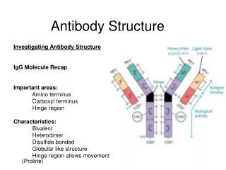
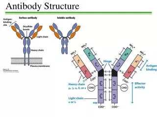
D2 Antibody structure 1.Light and heavy chains Each IgG antibody molecule consists of four polypeptide chains (two identical light chains and two identical heavy chains joined by disulfide bonds) and has two antigen-binding sites (i.e. is bivalent).
Variable and constant regions • Each light chain and each heavy chain consists of a variable region and a constant region. Variability in the variable regions is largely confined to three hypervariable regions ;the remaining parts of the variable regions are far less variable and are called the framework regions .
Antibody domains • Each light chain folds into two domains ,one for the variable region and one for the constant region . Each IgG heavy chain folds into four domains, one for the variable region and three in the constant region .
Fab and Fc fragments • Papain IgG into two Fab fragments (each of which has an antigen for complement activation and phagocytosis ).Pepsin digests IgG to release an F(ab`) 2fragment that has two antigen-binding sites.
Five classes of immunoglobulins • Human immmunoglobulins exist as IgA、IgD、IgE、IgG and IgM classes which contain αδεγandμ heavy chains,respectively. IgM is a pentamer that binds to invading microorganisms and activates complement killing of the cells and phagocytosis.
IgG • IgG is the main antibody found in the blood after antigen stimulation and also has the ability to cross the placenta .IgA mainly functions in body secretions .IgE provides immunity against some parasites but is also respomsible for the clinical symptoms of allergicteactions.The role of IgD is unknown. • All antibody molecules contain either kappa or lambda light chains .
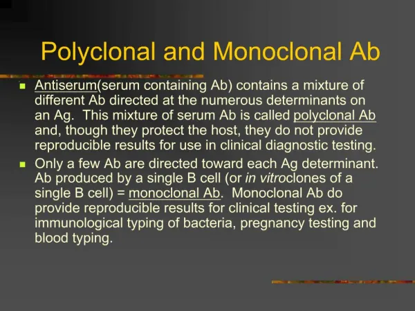
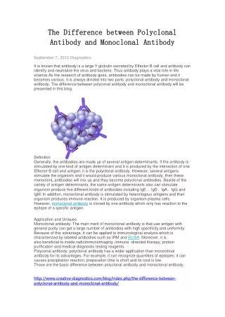
D3 Polyclonal and monoclonal antibodies • Polyclonal antibodies A preparation of antibody molecules that arises from several different clones of cells is called a polyclonal antibody .It is a mixture of antibody molecules that bind to different parts of the antigen and with different binding affinities.
Monoclonal antibodies • Antibody produced by a single clone of cells is a monoclonal antibody; all the antibody molecules are identical and bind to the same antigenic site with identical binding affinities. • Monoclonal antibodies can be ge-nerated in large amounts by creatint a cell fusion (called a hybridoma) between an antibody-producing cell and a mye-loma cell.
D4 Antibody synthesis • Somatic recombination No complete antibody gene exists in germ-line cells .The genes for light chains and heavy chains assemble by somatic recombination during blymphocyte maturation.
2. Recombination of light chain genes • In the germ-line ,each light chain gene exists as multiple V and J gene segments upstream of a single C gene segments. During B-lymphocyte differentiation , one V gene segment joins with one J gene segment (VJ joining) to assemble the complete light chain gene , usually by deletion of inter-vening DNA.
3. Recombination of heavy chain genes • Heavy chains are encoded by multiple V,J and D gene segments which lie upstream of a single copy of C gene segments for each of the constant regions of μ δ γ ε and α chains. During B-lymphocyte differentiation, a D gene segment joins a J segment (DJ joining)and then the recombined DJ joins a V gene segment(VDJ joining).
4. Class switching • A B lymphocyte can change the class of antibody being expressed by moving a new C gene segment into position after the recombined VDJ segment, deleting the intervening DNA. The new heavy chain has a different constant region but retains the same antigen-binding specificity of the previous heavy chain.
D5 Antibodies as tool • Immunolocalization methods Because of the high specificity of an antibody for its epitope, an antibody raised against a particular protein antigen can be used to determine the location of that antigen in a cell using immu-nofluorescence light microscopy or immuno-electron microscopy.
2. ELISA • ELISA can be used to quantify the amount of a specific protein antigen in a sample. The antibody is bound to an inert polymer support ,then exposed to reacts with the antigen at a different epitope is added.
Thesecondantibody • The second antibody used is one that has an enzyme attached to it that coverts a colorless or nonfluorescent substrate into a colored or fluorescent product. The amount of second antibody bound, and hence the amount of protein antigen present in the original sample, is determined by quan-tification of the intensity of color or fluorescence produced.
3. Western blotting • Protein samples are separated by one-dimensional SDS-PAGE of two-dime-nsional gel electrophoresis in polya-crylamide gels .The separated proteins are then transferred to a nitrocellulose or nylon sheet.
Autoradiography • This is incubated with specific antibody to the protein and then unbound antibody is washed away .Those proteins in the gel that bind the antibody are detected either by autoradiography (if the specific antibody was radiolabeled) or by using a second labeled antibody that binds to the primary antibody.
4. Immunoaffinity chromato-graphy • Immunoaffinity chromatography can be used to purify protein antigens by immo-bilizing the relevant antibodies on an inert matrix such as polysaccharide beads .When exposed to a protein mixture, only the protein recognized by that antibody will bind to the beads and can be eluted later in pure or almost pure form .Cells bearing the antigen on their surface can also be purified using a similar procedure.