Download
1 / 70
710 likes | 967 Vues
Human Anatomy, First Edition McKinley & O'Loughlin. Chapter 26 : Digestive System. General Structure and Functions of the Digestive System. Organs of the Digestive System to: Ingest the food. Transport the food. Digest the food into smaller usable components.

E N D
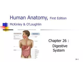
Human Anatomy, First EditionMcKinley & O'Loughlin Chapter 26 : Digestive System
General Structure and Functions of the Digestive System • Organs of the Digestive System to: • Ingest the food. • Transport the food. • Digest the food into smaller usable components. • Absorb the necessary nutrients into the bloodstream. • Expel the waste products from the body.
General Structure and Functions of the Digestive System • Composed of two separate categories of organs: • digestive organs • accessory digestive organs. • Digestive organs collectively make up the: • gastrointestinal (GI) tract. • Also called: • the digestive tract • alimentary canal.
General Structure and Functions of the Digestive System • The GI tract organs: • oral cavity • pharynx • esophagus • stomach • small intestine • large intestine • continuous tube • about 30 feet (9–10 meters) • from mouth to anus. • Smooth muscle in the wall • responsible for motility • pushes materials from one end to the other.
General Structure and Functions of the Digestive System • Accessory digestive organs: • do not form the GI tube • can develop as outgrowths • are connected to the GI tract (some by ducts) • Assist the GI tract in the digestion of food. • Include: • Teeth • Tongue • Salivary glands • Liver • Gallbladder • Pancreas
Digestive System Functions • Ingestion • Digestion: break down of large particles of food • mechanical digestion • chemical digestion • Propulsion • peristalsis • segmentation • Secretion: • digestive enzymes • hormones • Absorption: • from external environment into internal environment • across mucosa • Elimination of wastes (defecation)
Oral Cavity (mouth) • Entrance to the GI tract. • Initial site of digestion: • mechanical digestion (via mastication) • chemical digestion (via enzymes in saliva). • Bounded anteriorly by the teeth and lips • Bounded posteriorly by the oropharynx. • Superior boundary is formed by the hard and soft palates. • Floor, or inferior surface, of the oral cavity • the tongue • the mylohyoid muscle covered with mucosa.
Oral Cavity (mouth) • Two regions of the oral cavity • Vestibule is the space between the cheeks or lips and the gums. • Oral cavity proper. • The lateral walls are formed by the cheeks. • Contain buccinator muscles • Lips (labia). • Orbicularis oris muscle • Keratinized stratified squamous ET • Gingivae, or gums. • Dense regular CT • Nonkeratinized ET • Labial frenulum.
Palate • Hard palate • Anterior two-thirds of the palate • hard and bony • Soft palate • Posterior one-third • soft and muscular • primarily composed of skeletal muscle. • Extending inferiorly from the posterior part of the soft palate is the uvula. • When swallowing, the soft palate and the uvula elevate to close off the opening of the nasopharynx.
Palate • Fauces represent the opening between the oral cavity and the oropharynx. • Fauces are bounded by paired muscular folds: • glossopalatine arch (anterior fold) • pharyngopalatine arch (posterior fold) • Palatine tonsils are housed between the arches.
Tongue • An accessory digestive organ • Formed from: • skeletal muscle • covered with lightly keratinized stratified squamous epithelium. • Manipulates and mixes ingested materials during chewing • Forms the bolus. • a globular mass of partially digested material • Performs important functions in swallowing.
Tongue • Inferior surface of the tongue • attaches to the floor of the oral cavity • By the lingual frenulum. • Numerous small projections (papillae) cover the superior (dorsal) surface. • Posterior surface contains lingual tonsils. • Skeletal muscles move the tongue.
Salivary Glands • Collectively produce and secrete saliva. • a fluid that assists in the initial activities of digestion • Volume of saliva secreted daily ranges between 1.0 and 1.5 L. • Most is produced during mealtime • Smaller amounts are produced continuously to ensure that the oral cavity remains moist.
Salivary Glands • Components of saliva • Water: makes up 99% • Amylase: first step of chemical digestion • Lysozyme: antimicrobial • Functions • Moisten food • Food molecules into solution: taste • Form bolus: for swallowing • Cleanse oral cavity.
Salivary Glands • Three pairs of large, multicellular salivary glands: • parotid glands • submandibular glands • sublingual glands
The Parotid Glands • Largest salivary glands. • located anterior and inferior to the ear • partially overlying the masseter muscle. • Produce about 25–30% of saliva • conducted through the parotid duct to the oral cavity.
The Submandibular Glands • Inferior to the body of the mandible. • Produce most of the saliva (about 60–70%). • ducts opens through a papilla in the floor of the mouth • lateral to the the lingual frenulum.
The Sublingual Glands • Inferior to the tongue • internal to the oral cavity mucosa. • Each gland has multiple tiny sublingual ducts • open onto the inferior surface of the oral cavity • posterior to the submandibular duct papilla. • Contribute only about 3–5% of the total saliva.
Teeth • Collectively known as the dentition. • Responsible for mastication • first part of the mechanical digestion. • A tooth has: • exposed crown • constricted neck • one or more roots • Roots of the teeth fit into dental alveoli • are sockets within the alveolar processes • on both the maxillae and the mandible. • Collectively, the roots, the dental alveoli, and the periodontal ligament that binds the roots to the alveolar processes form a gomphosis joint.
Teeth • Two sets of teeth • 20 deciduous teeth, also called “milk teeth,” erupt between 6 months and 30 months after birth. • These teeth are eventually lost and replaced by 32 permanent teeth. • The more anteriorly placed permanent teeth tend to appear first, followed by the posteriorly placed teeth. • The last teeth to erupt are the third molars, often called “wisdom teeth,” in the late teens or early 20’s. • Often the jaw lacks space to accommodate these final molars, and they may either emerge only partially or grow at an angle and become impacted. • Impacted teeth cannot erupt properly because of the angle of their growth.
Pharynx • Review • Pharyngeal constrictors • Innervated by the vagus nerves
General arrangement of abdominal GI organs • Peritoneum • Parietal peritoneum • Visceral peritoneum • Peritoneal cavity • Intraperitoneal organs • Retroperitoneal organs
General arrangement of abdominal GI organs • Mesentaries • Double layered folds of peritoneum • Greater omentum • Lesser omentum • Mesentery proper • Suspends small intestine from posterior wall of abdomen • Mesocolon • Suspends large intestine • Peritoneal ligament • Peritoneum that attaches one organ to another
General Histology of GI Organs • from the esophagus through the large intestine • a tube • composed of four concentric layers called tunics. • From deep to superficial, these tunics are: • the mucosa • the submucosa • submucosal nerve plexus (Meissner plexus) • the muscularis • myenteric plexus (Auerbach plexus) • the adventitia or serosa
Esophagus • Tubular passageway • Pharynx to stomach • Bolus • About 25 cm in adult • Esophageal hiatus: through diaphragm • Histology • Mucosa: nonkeritinized stratified squamous ep. • Submucosa: thick, elastic fibers, mucous glands • Muscularis: inner circular, outer longitudinal • Both skeletal and smooth • Adventitia
Esophagus • Superior esophageal sphincter: • Skeletal muscle • Where pharynx and esophagus meet • Inferior esophageal sphincter • Also cardiac sphincter • Circular smooth muscle • Orifice between esophagus and stomach
Stomach • General • J-shaped • Functions • Digestion • Chemical • Mechanical • Results in chyme • Limited absorption
Stomach • Gross anatomy • Cardia • Cardiac orifice • Fundus • Body • Pylorus • Pyloric sphincter • Pyloric orifice • Greater curvature • Greater omentum • Lesser curvature • Lesser omemtum • Gastric folds (rugae)
Stomach • Histology • Mucosa: simple columnar • Gastric pits • Gastric glands • Muscularis • 3 layers • Inner oblique • Middle circular • Outer longitudinal
Small Intestine • Finishes chemical digestion • Responsible for absorbing most of the nutrients. • Ingested nutrients spend at least 12 hours in the small intestine. • thin-walled tube • about 6 meters (20 feet) in length. • coiled • Extends from the pylorus of the stomach to the cecum of the large intestine • occupies a significant portion of the abdominal cavity.