Download
1 / 26
260 likes | 462 Vues
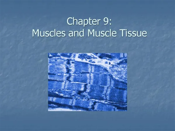
Muscles and Muscle Tissue . Muscular Tissue. Characteristics Excitability: tissue is able to respond to a stimulus Extensibility: tissue is able to be stretched Elasticity: tissue is able to return to normal after stretching. Contractibility: tissue is able to shorten and thicken .

E N D
Muscular Tissue • Characteristics • Excitability: tissue is able to respond to a stimulus • Extensibility: tissue is able to be stretched • Elasticity: tissue is able to return to normal after stretching. • Contractibility: tissue is able to shorten and thicken
Functions: • Motion- both reflex and voluntary • Maintenance of posture and organ volume • Thermogenesis- heat production • Joint Stability- allows for motion and maintenance of posture.
Muscle Fiber Types 3 Types of Muscle- 1. Smooth 2. Cardiac 3. Skeletal/Striated
Skeletal Muscle • Fascia: sheets or broad bands of fibrous connective tissue located beneath skin or around muscle or body organs. • Categories of Fascia: • Superficial Fascia- Structure: contains adipose, blood vessels, nerves Functions: stores fat, provides insulation, protects from injury
Categories of Fascia (cont): • Deep Fascia: no fat • Surrounds muscle groups and divides muscle groups (allows individual and independent movement) • Arrangement: • Endomysium: fascia which surrounds each muscle fiber • Perimysium: fascia which surrounds a bundle of muscle fibers (fascicle) • Epimysium: connective tissue which wraps the entire muscle
Muscle Attachments • Direct (Fleshy) attachments- perimysium is fused to peristeum of bone • Indirect attachment- the muscle fascia extends beyond the muscle and attaches as a rope-like tendon or aponeurosis. - Muscles are anchored to one another
Arrangement of Fascicles: • Parallel- run with the longitudinal axis of the muscle • Strap like- belly is uniform • Fusiform- belly is enlarged
Arrangement of Fascicles • Pennate- arranged obliquely to the bone A. Unipennate- one side B. Bipennate- both sides • Convergent broad at origin, narrows to a single tendon. • Circular- (sphincter) control diameter of an opening eye squinting, lip puckering
Muscle Cell Structure • 1. Sarcolema- plasma membrane surface • Sarcoplasm: cytoplasm and glycogen • 20 or more nuclei • Proteins: myoglobin- binds oxygen (red protein) • Usual organelles: (mitochondria) • Sarcoplasmic Reticulum: run parallel with myofibrils, densly packed
Myofibrils: protiens • A. Myosin: thick filaments • 2 globular heads • B. Actin: thin filaments • Active sights where globular heads of myosin attach • C. Tropomyosin: rod shaped spirals around actin, stiffens • D. Troponin: 3 polypeptides • T-Tubules- extensions of sarcolema that extend up into the sarcoplasm. • Conducts nerve impulses deep into muscle cells • Transports and regulates glucose, oxygen, calcium
Arrangement of Myofibrils • Sarcomere: functional unit of muscle
Sliding Filament Theory 1. Nerve impulse stimulates muscles. T-tubules release Calcium ion. Contraction is triggered. • Cross-bridge attachment- globular heads of myosin attach to active sites on actin. • Power stroke- as myosin binds and pivots: high energy to low energy actin slides over to the center of the sarcomere • Cross bridge detachment: requires ATP • Cocking of Myosin Heads: (rearmed) * Rigomortis: peaks at 12 hours
http://highered.mcgraw-hill.com/sites/0072437316/student_view0/chapter42/animations.html#http://highered.mcgraw-hill.com/sites/0072437316/student_view0/chapter42/animations.html#
All or None Phenomenon • When stimulated a muscle fiber contracts to its fullest capacity
Neuromuscular Junction: • Area where axonal end of a neruon fomrs a synaptic cleft with a muscle fiber. • Acetylcholine- neurotransmitter diffueses across the synaptic cleft and binds to receptors on the sarcolema, then destroyed by cholinesterase. • 1 impusle causes 1 contraction
Muscle Tone- • Partial contraction of a muscle fiber due to use • Awake- highest • Asleep- lower • unconscious- absent • Twitch: single muscle contraction (1/10 of a contraction) • Tetanus- prolonged muscle contraction required to do work. • Myogram- record of a muscle contraction
Muscle contraction • Isotonic contraction: muscle shortens, movement occurs. • Isometric contraction: muscle tenses but does not shorten
Muscle fiber types • Type I (red, slow twitch) postural muscles long distance • Large amounts of myoglobin • Many mitochondria • Many blood capillaries • Very good at generating ATP • Split ATP slowly – very resistant to fatigue
Type II A- intermediate, fast twitch fast oxidative - very large amounts of myoglobin - many mitochondria - very large supply of blood capillaries - split ATP very rapidly - very quick contraction velocity - less resistant to fatigue (sprinters, arms)
Type II B- white fast twitch, fast glycolytic - low myoglobin content - few mitochondria - few capillaries - large amounts of glycogen - geared for anaerobic respiration - split ATP rapidly - very little resistance to fatigue
Oxygen Debt the additional oxygen needed after exercise to return to homeostasis hyperventilation: to get rid of oxygen debt