Download
1 / 174
1.74k likes | 1.77k Vues
Learn about the types and functions of muscle tissues, along with their gross anatomy, connective tissue sheaths, and more.

E N D
Muscles • The most distinguishing functional characteristic of muscles is their ability to transform chemical energy (ATP) into directed mechanical energy • In doing this, they become capable of exerting force • Terminology: • Skeletal and smooth muscle cells (but not cardiac muscle cells) are elongated and, for this reason, are called muscle fibers • Muscle contraction depends on two kinds of myofilaments, which are the muscle equivalents of the actin-or-myosin-containing cellular microfilaments • Proteins that play a role in motility and shape changes in virtually every cell in the body • Prefixes myo or mys (both are word roots meaning “muscle”) • Prefix sarco (flesh), the reference is to muscle • Example: • Sarcolemma: plasma membrane of muscle cell • Sarcoplasm: muscle fiber cytoplasm
Types of Muscle Tissue • Skeletal muscle is associated with the bony skeleton, and consists of large cells that bear striations and are controlled voluntarily • Skeletal muscle fibers are the longest muscle cells • Only muscle cells subject to conscious control • Cardiac muscle occurs only in the heart, and consists of small cells that are striated and under involuntary control • Smooth muscle is found in the walls of hollow visceral organs (stomach, urinary bladder, and respiratory system), and consists of small elongated cells (fibers) that are not striated and are under involuntary control
Functional Characteristics of Muscle Tissue • Excitability, or irritability, is the ability to receive and respond to a stimulus • The stimulus is usually a chemical—for example, a neurotransmitter released by a nerve cell, or a local change on pH • Response is generation of an electrical impulse that passes along the sarcolemma (plasma membrane) of the muscle cell and causes the cell to contract • Contractility is the ability to contract (shorten) forcibly when stimulated • Extensibility is the ability to be stretched or extended • Muscle fibers (cells) shorten when contracted, but they can be stretched, even beyond their resting length, when relaxed • Elasticity is the ability of a muscle fiber (cell) to resume to its original length (recoil) after being stretched
Muscle Functions • Muscles produce movement by acting on the bones of the skeleton, pumping blood, or propelling substances throughout hollow organ systems (digestive, circulatory, urinary, reproductive systems) • Muscles aid in maintaining posture by adjusting the position of the body with respect to gravity • Muscles stabilize joints by exerting tension around the joint • Muscles generate heat (as they contract) as a function of their cellular metabolic processes • Important in maintaining normal body temperature
Gross Anatomy of Skeletal Muscle • Each muscle has a nerve and blood supply that allows neural control and ensures adequate nutrient delivery and waste removal • In general each muscle is served by one nerve, an artery, and by one or more veins • All of which enter or exit near the central part of the muscle and branch profusely through its connective tissue sheaths • Muscle capillaries, the smallest of the body’s blood vessels, are long and winding and have numerous cross-links, features that accommodate changes in muscle length • They straighten when the muscle is stretched and contort when the muscle contracts
Connective Tissue Sheaths • In an intact muscle, the individual muscle fibers (cells) are wrapped and held together by several different connective tissue sheaths (coverings) • Together these connective tissue sheaths support each cell and reinforce the muscle as a whole: • Endomysium surrounds each muscle fiber (cell) • Perimysium surrounds groups of muscle fibers • Epimysium surrounds whole muscle
Endomysium • A fine sheath of connective tissue consisting mostly of reticular fibers that surround each individual muscle fiber (cell)
Perimysium and Fascicles • Within each skeletal muscle, the endomysium-wrapped muscle fibers are grouped into fascicles that resemble bundles of sticks • Surrounding each fascicle is a layer of fibrous connective tissue called perimysium
Epimysium • An “overcoat” of dense irregular connective tissue surrounds the whole muscle • Sometimes the epimysium blends with the deep fascia that lies between neighboring muscles or the superficial fascia deep to the skin
Connective Tissue Sheaths of Skeletal Muscle • All of these connective tissue sheaths are continuous with one another as well as with the tendons that join muscles to bones • Therefore, when muscle fibers contract, they pull on these sheaths, which in turn transmit the pulling force to the bone to be moved • They also contribute to the natural elasticity of muscle tissue, and for this reason these elements are sometimes referred to collectively as the “series elastic components” • They also provide entry and exit routes for the blood vessels and nerve fibers that serve the muscle
Attachments • Span joints and cause movement to occur from the movable bone (the muscle’s insertion)toward the less movable bone (the muscle’s origin) • In the muscles of the limbs, the origin typically lies proximal to the insertion
Attachments • Muscle attachment may be direct or indirect • Direct: fleshy attachment • The epimysium of the muscle is fusedto theperiosteum of a bone or perichondrium of a cartilage • Indirect: • Much more common because of their durability and small size • The muscle’s connective tissue wrappings extend beyond the muscle either as a ropelike tendon or as a sheet-like aponeurosis (flat fibrous sheet of connective tissue that attaches muscle to bone or other tissues—may sometimes serve as a fascia) • Tendons are mostly tough collagenic fibers: • They cross rough bony projections that would tear apart the more delicate muscle tissues • Because of their relatively small size, more tendons than fleshy muscles can pass over a joint—thus, tendons also conserve space
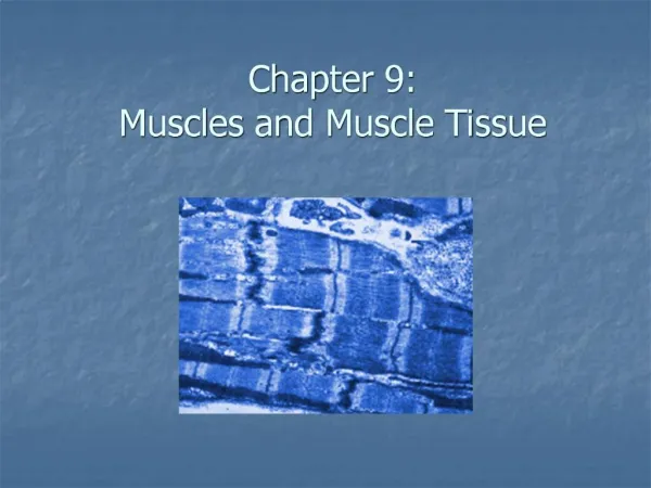
Microscopic Anatomy of a Skeletal Muscle Fiber • Skeletal muscle fibers are long cylindrical cells with multiple nuclei beneath the sarcolemma • Skeletal muscle fibers are huge cells • Their diameter typically ranges from 10 to 100 um—up to ten times that of an average body cell—and their length is phenomenal, some up to 30 cm long
Microscopic Anatomy of a Skeletal Muscle Fiber • Sarcoplasm of a muscle fiber is similar to the cytoplasm of other cells, but it contains unusually large amounts of glycosomes (granules of stored glycogen) and substantial amounts of an oxygen-binding protein called myoglobin • Myoglobin, a red pigment that stores oxygen, is similar to hemoglobin, the pigment that transports oxygen in blood
Microscopic Anatomy of a Skeletal Muscle Fiber • The usual organelles are present, along with some that are highly modified in muscle fibers: myofibrils and the sarcoplasmic reticulum • T tubules are unique modification of the sarcolemma
Myofibrils (b) • Each muscle fiber contains a large number of rodlike myofibrils that run parallel to it length • Densely packed in the fiber that mitochondria and other organelles appear to be squeezed between them • Myofibrils account for roughly 80% of cellular volume, and contain the contractile elements of the muscle cell
Striations (c) • Due to a repeating series of dark A bands and light I bands • A band has a lighter stripe in its midsection called the H zone • Visible only in relaxed muscle fibers • Each H zone is bisected vertically by a dark line called the M line • The I bands also have a midline interruption, a darker area called the Z disc
Striations (c)Sarcomere • Region of a myofibril between two successive Z dics, that is, it contains an A band flanked by half an I band at each end • Smallest contractile unit of a muscle fiber • Functional units of skeletal muscles
Myofilaments (d) • If we examine the banding pattern of a myofibril at the molecular level, we see that it arises from an orderly arrangement of two types of even smaller structures, called myofilaments or filaments, within the sarcomeres • Myofilaments make up the myofibrils, and consist of thick and thin filaments
Myofilaments (d) • Central thick filaments extend the entire length of the A band • The more lateral thin filaments extend across the I band and partway into the A band • The Z disc composed of the protein nebulin anchors the thin filaments and connects each myofibril to the next throughout the width of the muscle cell
Myofilaments (d) • H zone of the A band appears less dense because the thin filaments do not extend into this region • M line in the center of the H zone is slightly darker because of the presence of fine protein strands that hold adjacent thick filaments together
Ultrastructure and Molecular Composition of the Myofilaments • There are two types of myofilaments in muscle cells: • (a): thick filaments are composed primarily of bundles of protein myosin • Each myosin molecule has a rodlike tail terminating in two globular heads and a tail of two interwoven heavy polypeptide chains • The heads link the thick and thin filaments together (cross bridges) during contraction
Ultrastructure and Molecular Composition of the Myofilaments • (b+d): Each thick filament contains about 200 myosin molecules bundled together with their tails forming the central part of the thick filament and their heads facing outward and in opposite direction at each end • Besides bearing actin binding sites, the heads contain ATP binding sites and ATPase enzymes that split ATP to generate energy for muscle contraction
Ultrastructure and Molecular Composition of the Myofilaments • (c): Thin filaments are composed of strands of actin • The backbone of each thin filament appears to be formed by an actin filament that coils back on itself, forming a helical structure that looks like a twisted double strand of pearls
Ultrastructure and Molecular Composition of the Myofilaments • Several regulatory proteins are also present in the thin filament • Two strands of tropomyosin, a rod-shaped protein, spiral about the actin core and help stiffen it • The other major protein in the thin filament, troponin, is a three-polypeptide complex • One of these polypeptides (TnI) is an inhibitory subunit that binds to actin • Another (TnT) binds to tropomyosin and helps position it on actin • The third (TnC) binds calcium ions • Both tropomyosin and troponin are regulatory proteins present in thin filaments and help control the myosin-actin interactions involved in contraction
Skeletal Muscle Fibers (cells) • Contain two sets of intracellular tubules that participate in regulation of muscle contraction: • 1. Sarcoplasmic reticulum • 2. T tubules
Sarcoplasmic (SR) • Is a smooth endoplasmic reticulum surrounding each myofibril • Major role is to regulate intracellular levels of ionic calcium: • It stores calcium and releases it on demand when the muscle fiber is stimulated to contract
Relationship of the Sarcoplasmic Reticulum and T tubules to the Myofibrils of Skeletal Muscle
T Tubules • Are infoldings of the sarcolemma that penetrate into the cell interior to form an elongated tube • Muscle contraction is ultimately controlled by nerve-initiated electrical impulses that travel along the sarcolemma • Because T tubules are continuations of the sarcolemma, they can conduct impulses to the deepest regions of the muscle cell and to every sarcomere • These impulses signal for the release of calcium from the adjacent terminal cisternae
Relationship of the Sarcoplasmic Reticulum and T tubules to the Myofibrils of Skeletal Muscle
Sliding Filament Model of Contraction • Sliding Filament Theory of Contraction: • States that during contraction the thin filaments slide past the thick ones so that the actin and myosin filaments overlap to a greater degree • Overlap between the myofilaments increases and the sarcomere and the sarcomere shortens
Sliding Filament Model of Contraction • (1): Relaxed State: • In a relaxed muscle fiber (cell), the thick and thin filaments overlap only slightly • When muscle fibers are stimulated by the nervous system, the cross bridges latch on to myosin binding sites on actin in the thin filaments, and the sliding begins