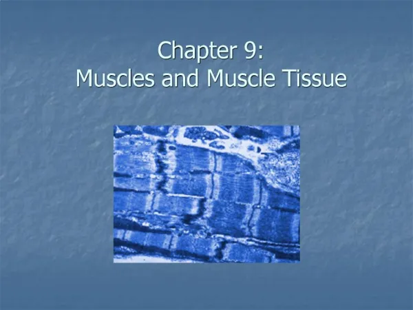
Chapter 9 Muscles and Muscle Tissue
190 likes | 541 Vues
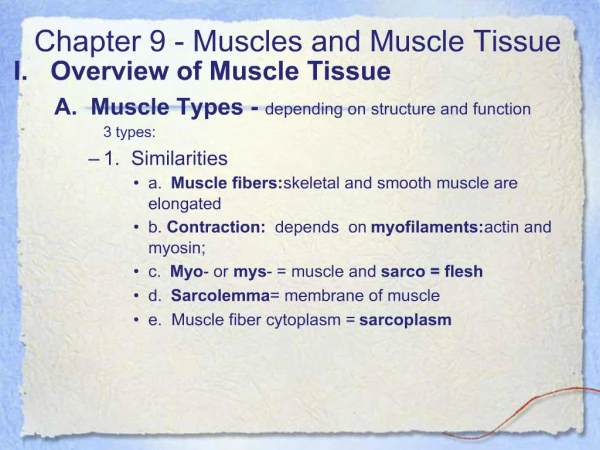
Chapter 9 Muscles and Muscle Tissue. Sarcomere and Contraction. Skeletal Muscle Fibers – Regions of the Sarcomere. Sarcomere the smallest functional unit of the muscle fiber. Interactions between the thick and thin filaments of sarcomeres are responsible for muscle contraction.

Chapter 9 Muscles and Muscle Tissue
E N D
Presentation Transcript
Chapter 9Muscles and Muscle Tissue Sarcomere and Contraction
Skeletal Muscle Fibers – Regions of the Sarcomere • Sarcomere • the smallest functional unit of the muscle fiber. • Interactions between the thick and thin filaments of sarcomeres are responsible for muscle contraction
Skeletal Muscle Fibers – Regions of the Sarcomere • A band – Dark band; mainly thick filaments • I band – Light band; mainly thin filaments • Note: In skeletal muscle fibers, these bands are almost perfectly aligned giving the striped appearance known as striations.
Skeletal Muscle Fibers – Regions of the Sarcomere • M line – Central portion of each thick filament • H zone/line – lighter region on either side of the M line. Only seen in resting muscle fibers
Skeletal Muscle Fibers – Regions of the Sarcomere • Zone of overlap – where the thin filaments are found between thick filaments • Z line- mark the boundary lines between adjacent sarcomeres (midline interruption of the I band)
Sliding Filament Theory • Myosin heads attach to and “walk” along the thin filaments pulling the thin filaments towards the M line thin filaments are sliding toward the center of the sarcomere
Sliding Filament Theory • Thin filaments slide inwards: • H band gets smaller (disappears) • I band gets smaller (disappears) • Z lines move closer together • A band width remains constant • Sarcomere shortens
Skeletal Muscle Fiber Contraction • Nerve impulse travels to muscle cell • The impulse travels in all directions over the muscle cell and travels through the t tubules.
Skeletal Muscle Fiber Contraction • Impulse reaches the sarcoplasmic reticulum • Calcium ions diffuse from the SER into the sarcoplasm • Ca+2 bind to troponin (on actin) changes shape
Skeletal Muscle Fiber Contraction 6. The active site on actin is exposed 7. Actin and myosin filaments form a cross-bridge 8. Release the phosphate group (from hydrolyzed ATP)
Skeletal Muscle Fiber Contraction 9. Myosin heads pull actin filaments inward myosin head falls down 10. ADP is released. 11. ATP Binds to myosin
Skeletal Muscle Fiber Contraction 12. Cross-bridge (myosin)releases 13. ATP on the myosin is hydrolized to ADP and a phosphate group 14. Myosin head lifts to attach to actin
Skeletal Muscle Fiber Contraction 15. Continues until Calcium supply is cut off 16. The muscle fiber shortens as contraction occurs
Skeletal Muscle Fiber Relaxation • Calcium ions are transported into SER • ATP causes cross-bridge between actin and myosin filaments to break apart • The binding sites on actin are blocked (there is no calcium)
Skeletal Muscle Fiber Relaxation • Muscle fiber releases. • ATP breakdown “cocks” myosin cross-bridges. 6. Muscle fiber is ready for further stimulation
Muscle Contraction Videos • http://www.youtube.com/watch?v=83yNoEJyP6g • http://www.youtube.com/watch?v=WRxsOMenNQM • Day 2 • http://www.youtube.com/watch?v=mWPmUqRZYls • http://www.youtube.com/watch?NR=1&v=CbfK1Gi-aCk
http://www.youtube.com/watch?v=0kFmbrRJq4w • http://www.youtube.com/watch?v=EdHzKYDxrKc • http://www.youtube.com/watch?v=mWPmUqRZYls • http://www.youtube.com/watch?v=kvMFdNw35L0 • http://www.youtube.com/watch?v=Vlchs4omFDM
Rigor Mortis • After death, calcium ions start to leak out of SER • Causes muscle contraction cycle to start • ATP synthesis has stopped cell runs out of ATP • Crossbridges can’t break apart muscles in a state of contraction • Starts about 3-4 hours after death • Lasts about 24 hours (crossbridges are broken down)