Acute Leukemia
Hematologic Malignancies. Acute Leukemia. Definition. Leuko = white Emia = blood


Acute Leukemia
E N D
Presentation Transcript
Hematologic Malignancies Acute Leukemia
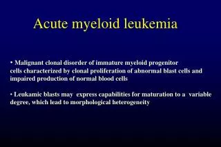
Definition Leuko = white Emia = blood Acute Leukemia: is a stem cell malignant disorder characterized by abnormal proliferation of malignant clone in uncontrolled fashion. It is a bone marrow based malignancy where expansion of the malignant clone interfere with normal BM function
Types Acute leukemia: runs a rapid and explosive course. Chronic Leukemia: runs a more protracted and less severe course.
Etiology Exact cause: Still unknown? Possible etiologic associations: 1- Radiation- Ionizing radiation exposure increases incidence of AL. Evidence from atomic bomb survivors in Japan & ankylosingspondylitis patients were treated with radiotherapy. No risk from diagnostic radiation in patients & medical personnel.
2- V iruses: Oncogenic viruses HTLV-I associated with ATLL HTLV-II causes in experimental animals a syndrome similar to Hairy cell Leukemia
3- Genetics: Increased AL in hereditary diseases with chromosomal instability AR- Ataxia-telangiectasia - Bloom syndrome - Fanconi Anemia X-linked infantile agammaglobulinemia Others- Down’s synd, Turner, Kleinfelter
Inv 16 – AML M4 t( 9;22) Ph + ALL, AML, CML t(15;17) – AML M3 t(10;14)(q24;q11) – ALL t(8;14)(q24;q32) Hyperdiploidy
4- Chemicals: Benzene, insecticides, pesticides, herbicides Drugs: Alkylating agents- CTx, chlorambucil, melphalan Nitrosoureas, etoposide Bimolane – used for psoriasis
5- Chronic BM stem cell disease e.g. PNH,AA,MDS ....etc Others- Potent EM fields - smoking
Epidemiology More in developed countries Male > female ALL in children > adults
classification FAB (Frech-American-British) 1- Acute lymphoblastic Leukemia (ALL) L1 in children L2 in adults L3 Burkitt-lymphoma-like 2- Acute myeloblasticLeukemia (AML)
AML M0 – acute undifferentiated leukemia M1 - AML with minimal differentiation M2- AML with differentiation M3- Acute promyelocyticLeukemia M4 – Acute myelomonoblasticleuk M4E – with peripheral eosinophilia M5- Acute monoblasticleukemia M6- Acute erythroleukemia M7- Acute megakaryoblasticleukemia
Clinical Manifestations Dx suspected in presence of Triad ( Anemia, fever & bleeding) due to BM occupation blasts Anemia- pallor, palpitation, easy fatigue, anorexia, SOB, Chest pain, dizziness Fever- due to neutropenia & infection ( G-ve bacilli, coagulase –ve staph, viruses, fungi...) Bleeding- hemorrhage into skin (petechiae, purpura, ecchymoses, bruises), gum, mucosae
Bone pain may be so severe that mimic JCA Hepatosplenomegaly – ALL > AML Lymphadenopathy – ALL > AML Peculiar features- Soft tissues myeloblastoma (Chloroma) – AML Cutaneous infiltration – M4,M5 Meningeal infiltration- ALL< M4,M5 Testicular infiltration- ALL Gum hypertrophy- M4,M5 Lactic acidosis- L3 Prolonged prodromal phase- M6 BM fibrosis- M7 Leukostasis- AML
Lab Dx CBC- Anemia- Hb low, platelets low - WBC may be increased (if >50000 risk of hyperleukocytosis), may be normal, may be decreased. - blasts are seen. If scanty (subleukemic). If absent ( Aleukemicleukemia) Schistocytes- DIC (M3)
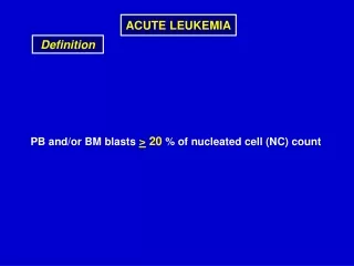
BMA&B- Hypercellularhyperplastic active Blasts > 30% of nucleated cells Decreased megakaryocytes BM fibrosis (M7) reticulin stain
Chemistry S uric acid increased SLDH increased Serum lysozyme increased
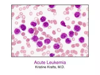
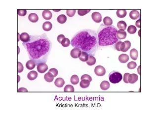
Typing Light microscope – using conventional stains detect blasts shape, nucleus size & shape Blasts in AML look larger with large nucleus, open chromatin with cytoplasmic inclusion bodies (Auer’s rods) Blasts in L1 look homogeneous, small size, high N/C ratio L2 larger, heterogeneous, low N/c ratio L3 large, homogeneous, pale vacuolated cytoplasm, similar to Burkitt NHL cells
Special Stains Sudan Black (SB-B) +ve AML MPO stain +ve AML PAS stain +ve M4,M5, M6, L3 tdT +ve ALL L1&L2 Electron microscopy – detect minimal MPO positivity in perinuclear space, Golgi, endoplasmic reticulum
Immunophenotyping Utilizing Cluster differentiation (CD) Flow cytometry or slide agglutination CD10 – L1 CD3 – T cell ALL CD19- B lineage ALL CD33- myeloid marker CD13- = = = CD41,42 – platelet
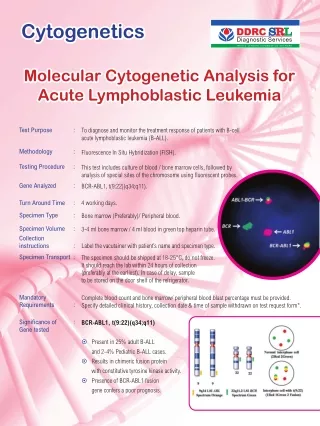
Karyotyping & Cytogenetics Chromosomal number & abnormalities Hyperdiploidy , hypodiploidy Dletion, inversion, mutation, translocation Philadelphia chromosome t(9;22)
Molecular Biology Using PCR & FISH BCR/ABL hybrid gene C-myc mutation PML mutation MLL mutation RARA gene over expression FLT3 gene mutation
DDx ALL- Acute MyelogenousLeukemiaLymphoma, B-CellLymphoma, High-Grade Malignant ImmunoblasticLymphoma, Mantle CellLymphoma, Non-HodgkinAcute biphenotypicleukemiaNK-cell leukemia
DDx AML- Acute Lymphoblastic LeukemiaLymphoma, B-CellAgnogenic Myeloid Metaplasia With MyelofibrosisLymphoma, LymphoblasticAgranulocytosisMyelodysplastic SyndromeMyelophthisic AnemiaAplastic AnemiaChronic MyelogenousLeukemia