Restriction Fragments and Mapping
60 likes | 736 Vues
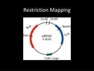
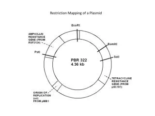
Restriction Fragments and Mapping. Restriction Fragment Analysis System used to compare the genes and DNA sequences between individuals in a population. can be used to identify heterozygous carriers of mutant alleles Uses restriction enzymes to digest (break apart) DNA into shorter segments.

Restriction Fragments and Mapping
E N D
Presentation Transcript
Restriction Fragments and Mapping • Restriction Fragment Analysis • System used to compare the genes and DNA sequences between individuals in a population. • can be used to identify heterozygous carriers of mutant alleles • Uses restriction enzymes to digest (break apart) DNA into shorter segments. • DNA segments may differ in length based on the mutations present in genes • restriction enzymes are specific for nucleotide sequences • a change in the sequence may cause an enzyme not to make a cut resulting in a larger segment • RFLPs (restriction length fragment polymorphisms) present • non-coding sections of DNA used to identify different relatedness of individuals in a population • can be used to construct linkage maps
Southern Blotting • Segments can then be compared to find the differences by gel electrophoresis - southern blotting • gel electrophoresis takes advantage of the overall negative charge associated with DNA molecules • DNA digests are put into wells (holes in the gel) and guided through the gel by an applied electrical current • gel acts as a molecular sieve(filter) allowing the smaller molecules to travel the furthest in a given amount of time • fluorescent or radioactive markers are then added to the gel to elucidate the bands present
Linkage mapping by FISH (fluorescent in situ Hybridization) • Fluorescent probes create a map of whole chromosomes as they hybridize with them. • the distance between the individual fluorescent probes creates a map of the chromosome • The physical map is then constructed as DNA digests are compared to find areas of overlap • accomplished through cloning with YACs & BACs • Once reconstructed the individual fragments can be fed through a sequencing machine to establish the nucleotide sequence
DNA Sequencing: The human Genome Project that spanned from 1993 to 2003 pushed the development of faster more efficient methods of DNA sequencing. • Dideoxy Chain-Termination Method • DNA to be sequenced is digested and amplified (phage vectors, YACs & BACs) • Cloned fragments are then sequenced using the dideoxy method • fragments are incubated in a test tube the following: • primers • DNA polymerase • deoxyribonucleotides (normal DNA components) • dideoxyribonucleotides • with the addition of a dDNA elongation of the growing strand is terminated • each different dDNA is fluorescently labeled with a different color • ddATP - green • ddCTP - blue • ddTTP - red • ddGTP - yellow • the result is many strands of different lengths with different colored termination points • the incubated fragments are then separated by weight and size through a polyacrylamide gel in a column (capillary tube) • finally a sequencing machine gives the sequence based on the weight and the end marker of each strand